블로그

  • Executive Function and Prefrontal Cortex in ADHD

    Executive Function and Prefrontal Cortex in ADHD

    Attention-deficit/hyperactivity disorder (ADHD) is a neurodevelopmental condition characterized by persistent patterns of inattention, hyperactivity, and impulsivity that interfere with daily functioning. ADHD executive function is described as a central mechanism underlying difficulties with planning, working memory, attention control, time management, and behavioral inhibition.

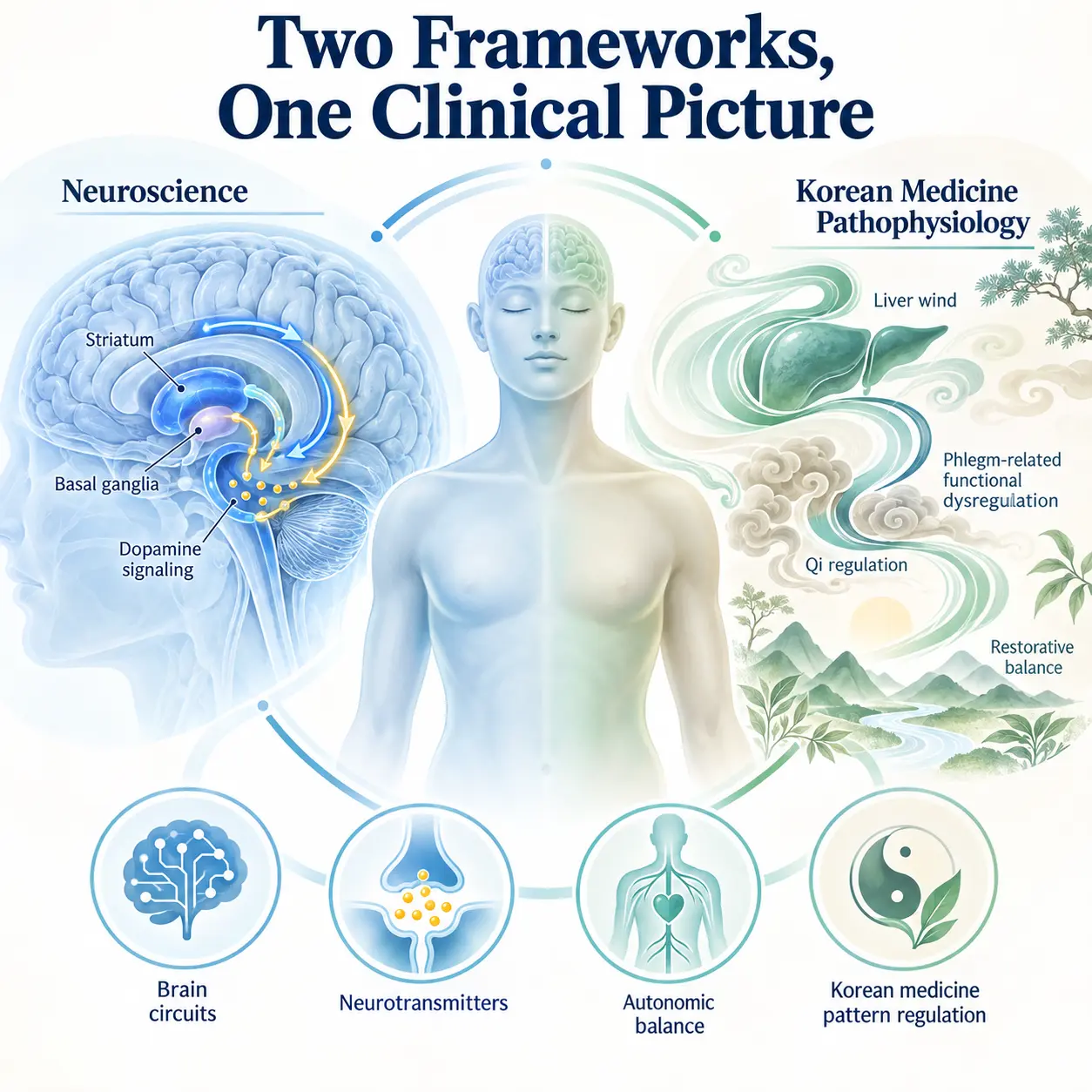
    Modern medicine explains ADHD as a disorder involving altered activity in the prefrontal cortex and fronto-striatal circuits, including the basal ganglia and striatum. In Korean medicine, the condition is understood as a disturbance of systemic regulation involving qi, blood, yin, and the functional stability of the mind.

    Korean Medicine Neuropsychiatry integrates these explanatory frameworks by examining how neural regulation and whole-body physiological balance are associated with attention and behavioral control. The Korean Society of Oriental Neuropsychiatry studies the relationship between psychiatric symptoms, nervous system function, and Korean medicine pattern identification.

    Overview

    ADHD is considered to be a neurodevelopmental condition rather than a personality characteristic or a simple lack of effort. Its symptoms reflect differences in systems responsible for regulating attention, motivation, action selection, and inhibition. The concept of ADHD executive function helps explain why a person may understand what needs to be done but still experience difficulty beginning, organizing, or completing the task.

    Executive function is described as a group of higher-order cognitive abilities that coordinate goal-directed behavior. These abilities include working memory, cognitive flexibility, planning, prioritization, emotional regulation, self-monitoring, and response inhibition. Executive function does not refer to a single skill or one isolated brain area. It is associated with coordinated activity across multiple neural networks.

    The prefrontal cortex plays a major role in maintaining task goals, evaluating consequences, suppressing unsuitable responses, and adjusting behavior according to changing circumstances. ADHD executive function impairment may be explained by reduced efficiency in the interaction between the prefrontal cortex, basal ganglia, striatum, and other regions involved in attention and motivation.

    For a broader understanding of symptom patterns, see ADHD symptom overview.

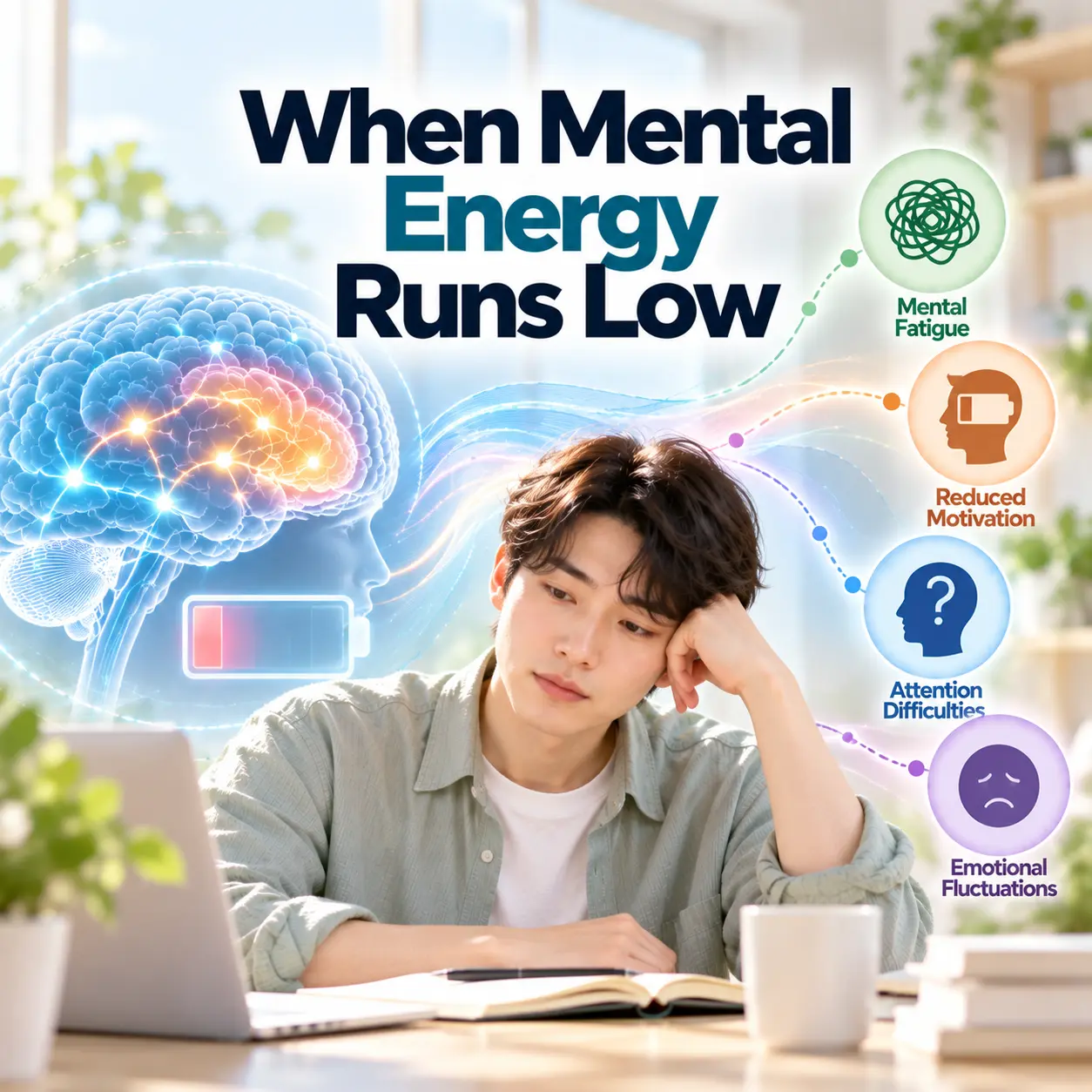
    Visualization of attention difficulty and distraction in ADHD executive function
    Attention difficulties in ADHD may involve problems selecting, sustaining, and shifting focus according to task demands.

    Clinical Features

    The core clinical features of ADHD include inattention, hyperactivity, and impulsivity. Their expression varies according to age, developmental stage, environment, and individual characteristics. Hyperactivity may appear as visible movement in childhood, while in adulthood it may be experienced as internal restlessness, impatience, or difficulty remaining mentally settled.

    Inattention is described as difficulty sustaining attention, particularly during repetitive or delayed-reward activities. A person may overlook details, lose necessary items, forget appointments, or move between tasks without completing them. These patterns are associated with ADHD executive function difficulties involving working memory, task sequencing, and self-monitoring.

    Impulsivity is understood as difficulty delaying a response long enough to consider its likely consequences. It may appear as interrupting, answering prematurely, making rapid decisions, or reacting strongly to immediate emotional cues. These behaviors may be explained by reduced inhibitory control and unstable regulation of reward-related signals.

    Common functional effects

    • Difficulty estimating how long a task will take
    • Frequent postponement of tasks requiring sustained concentration
    • Inconsistent performance despite adequate knowledge or ability
    • Problems organizing schedules, materials, or priorities
    • Emotional responses that occur faster than reflective control
    • Academic, occupational, relational, or household impairment

    Functional impairment is an important part of clinical evaluation. Occasional distraction alone does not establish ADHD. Symptoms are considered clinically significant when they are persistent, developmentally inappropriate, present across relevant contexts, and associated with meaningful impairment.

    Prefrontal cortex highlighted as the center of executive function in ADHD
    The prefrontal cortex supports working memory, planning, inhibitory control, and the regulation of goal-directed behavior.

    Etiology

    ADHD has a multifactorial etiology. It is associated with genetic vulnerability, differences in brain development, neurotransmitter regulation, stress exposure, sleep, and environmental demands. No single factor adequately explains every individual presentation.

    ADHD neurobiology is further described in academic research on brain function and attention regulation. National Institute of Mental Health ADHD overview provides additional scientific context.

    Prefrontal Cortex and Executive Regulation

    The prefrontal cortex is associated with the maintenance of goals, evaluation of competing information, inhibition of automatic responses, and adjustment of behavior. ADHD executive function impairment may be explained by reduced consistency in these regulatory processes rather than by a complete absence of executive ability.

    Performance may fluctuate according to novelty, urgency, personal interest, emotional significance, and the immediacy of reward. Consequently, an individual may concentrate intensely on one activity while finding it difficult to begin another activity of equal objective importance.

    Basal Ganglia, Striatum, and Fronto-Striatal Circuits

    The basal ganglia and striatum contribute to action selection, reward learning, habit formation, motor regulation, and motivational processing. Fronto-striatal circuits connect these regions with the prefrontal cortex and are considered to be central pathways in the neurobiology of ADHD.

    Altered fronto-striatal communication is associated with difficulty maintaining effort when rewards are delayed. It may also contribute to impulsive choices, variable reaction time, and difficulty suppressing responses that are immediately available but not aligned with a long-term goal.

    Fronto-striatal brain circuits involved in ADHD executive function
    Fronto-striatal circuits connect the prefrontal cortex with the striatum and basal ganglia to support cognitive and behavioral control.

    Dopamine and Norepinephrine

    Dopamine and norepinephrine are important neurotransmitters in attentional regulation, motivation, working memory, and response selection. Dopamine is associated with reward prediction, reinforcement, and motivational salience. Norepinephrine is associated with alertness, signal detection, and the maintenance of task-relevant information.

    Dysregulation of these neurotransmitter systems may reduce the signal-to-noise efficiency of prefrontal networks. ADHD executive function difficulties may therefore become more evident when a task requires sustained effort without immediate feedback or reward.

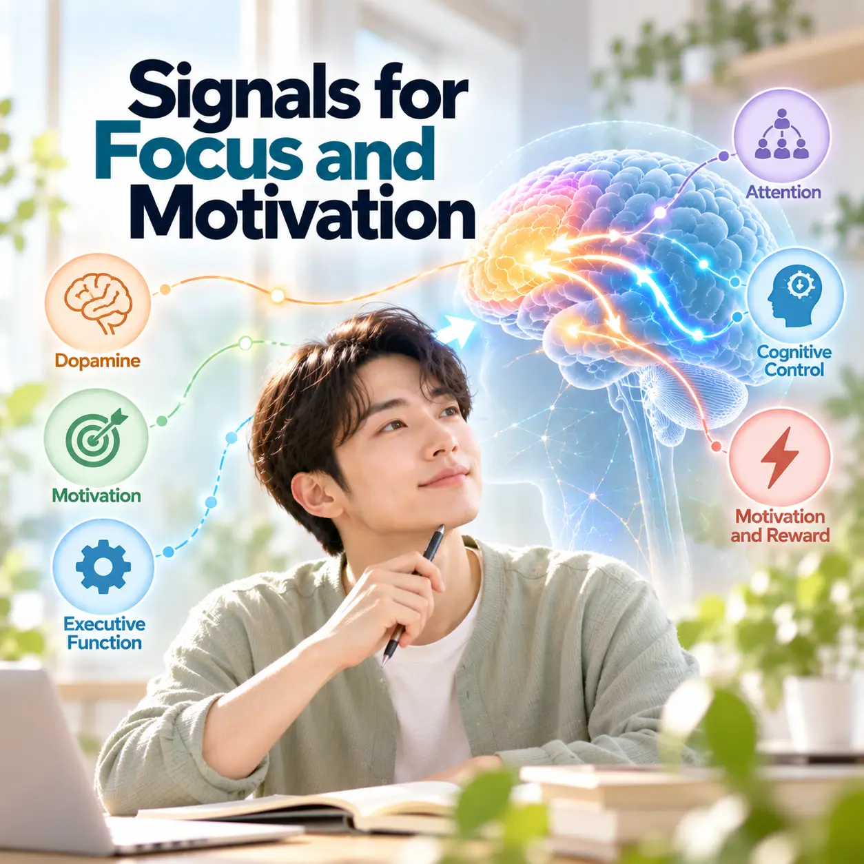
    Role of dopamine and norepinephrine in ADHD executive function
    Dopamine and norepinephrine contribute to attention, motivation, working memory, alertness, and executive control.

    Stress and Environmental Influences

    Environmental conditions do not provide a single explanation for ADHD, but they may influence symptom intensity and functional outcomes. Chronic stress, inconsistent routines, excessive cognitive demands, sleep disruption, and frequent interruptions can increase the burden on already vulnerable executive systems.

    Supportive structure, predictable routines, task segmentation, and reduced environmental distraction may improve functional performance. Such improvement does not mean that ADHD was caused only by the environment. It indicates that executive abilities are sensitive to the interaction between neurodevelopmental characteristics and contextual demands.

    Physiological System Changes

    ADHD is also associated with differences in arousal and physiological regulation. Effective attention requires neither maximum arousal nor complete relaxation. It depends on an appropriate level of activation that allows the brain to distinguish relevant signals from competing information.

    Autonomic Nervous System

    The autonomic nervous system regulates bodily functions such as heart rate, respiration, vascular tone, and physiological readiness. Its sympathetic branch supports mobilization, while its parasympathetic branch supports restoration and flexible regulation.

    Some individuals with ADHD may experience unstable arousal, shifting between underactivation and excessive activation. Underactivation may be associated with low task engagement or the need for stronger stimulation. Excessive activation may be associated with restlessness, emotional reactivity, or difficulty disengaging from immediate stimuli.

    Autonomic nervous system regulation related to ADHD symptoms
    Balanced autonomic regulation supports flexible shifts between activation, attention, and recovery.

    Stress Hormones and Body Responses

    The hypothalamic-pituitary-adrenal axis coordinates hormonal responses to stress, including cortisol release. Repeated or prolonged stress may affect sleep, emotional regulation, working memory, and the ability to maintain attention under pressure.

    Physiological responses may include muscle tension, altered breathing, fatigue, sleep irregularity, gastrointestinal discomfort, or heightened sensitivity to stimulation. These responses are not specific to ADHD, but they can increase the functional burden of ADHD executive function impairment.

    From an integrated perspective, executive control is understood as dependent on both neural circuitry and the physiological state of the body. When stress regulation and autonomic balance are unstable, the prefrontal cortex may have greater difficulty maintaining reflective control over immediate responses.

    Korean Medicine Pathophysiology

    Korean medicine describes ADHD-related symptoms through functional patterns rather than through one fixed disease mechanism. Pattern identification examines the relationship among attention, activity, emotion, sleep, digestion, fatigue, and constitutional tendencies. These concepts provide an explanatory framework that can be discussed alongside neuroscience without treating either framework as merely secondary.

    Qi and Blood Deficiency

    Qi and blood deficiency is described as insufficient functional support and nourishment for sustained mental activity. It may be linked to fatigue, weak concentration, forgetfulness, inconsistent task persistence, and reduced recovery after cognitive effort.

    Yin Deficiency

    Yin deficiency is understood as insufficient stabilizing and restorative capacity. It may be associated with internal restlessness, sleep disturbance, irritability, and difficulty settling attention after stimulation.

    Phlegm Accumulation

    Phlegm accumulation is described as an obstruction of clear mental processing. It may be linked to cognitive clouding, distractibility, heaviness, delayed task initiation, and difficulty organizing thoughts.

    Liver Qi Stagnation

    Liver qi stagnation is associated with constrained adaptation to stress and emotional pressure. It may be linked to irritability, frustration, variable concentration, impulsive reactions, and tension-related symptoms.

    These Korean medicine concepts may be functionally connected with nervous system regulation. Qi and blood deficiency may be discussed alongside reduced cognitive endurance, yin deficiency alongside impaired recovery and arousal regulation, phlegm accumulation alongside reduced mental clarity, and liver qi stagnation alongside stress-related autonomic activation.

    ADHD executive function impairment is therefore understood within Korean medicine as involving both insufficient support for stable cognition and difficulty regulating activation. This interpretation corresponds conceptually with neuroscience findings concerning prefrontal efficiency, fronto-striatal communication, autonomic balance, and stress-related changes in cognitive performance.

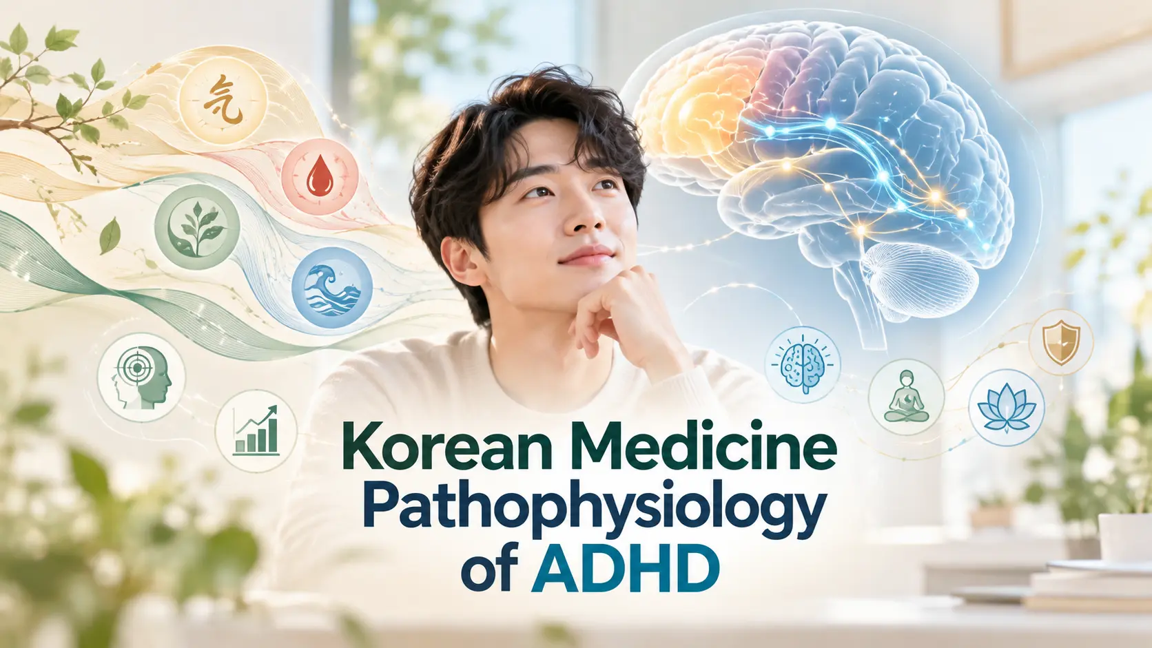
    Qi flow and systemic balance in Korean medicine interpretation of ADHD
    Korean medicine examines attention and behavioral regulation through functional relationships among qi, blood, yin, stress, and recovery.

    Treatment Perspective

    From the perspective of Korean Medicine Neuropsychiatry, treatment is understood as a process of supporting nervous system regulation, improving autonomic balance, and strengthening the recovery processes required for sustained cognitive control.

    The treatment perspective begins with an individualized assessment of symptom pattern, developmental history, sleep, emotional regulation, stress exposure, physical condition, and functional impairment. Because ADHD is a neurodevelopmental condition, treatment is not understood as correcting a personality flaw. It is directed toward reducing impairment and improving the consistency of self-regulation.

    Neuroscience-based approaches may focus on strengthening environmental structure, supporting prefrontal control, regulating dopamine and norepinephrine function, and reducing demands that unnecessarily overload working memory. Behavioral strategies may include task segmentation, external reminders, visual schedules, reduced distractions, and immediate feedback.

    Korean medicine pattern-based treatment is understood as addressing the functional imbalances associated with the individual presentation. Qi and blood deficiency may direct attention toward cognitive endurance and fatigue. Yin deficiency may direct attention toward restlessness, sleep, and recovery. Phlegm accumulation may direct attention toward mental clouding and slowed task initiation. Liver qi stagnation may direct attention toward stress reactivity and emotional constraint.

    The recovery process is considered to be multidimensional. Improvement may involve more stable sleep, better recognition of attention limits, reduced physiological overactivation, increased ability to pause before responding, and greater consistency in initiating and completing tasks. Changes in ADHD executive function are often more meaningful when evaluated through daily functioning rather than through concentration alone.

    Integrated view of brain function and body balance in ADHD executive function
    An integrated framework examines the relationship between prefrontal regulation, fronto-striatal circuits, autonomic balance, and systemic recovery.

    Summary

    ADHD is described as a neurodevelopmental condition involving persistent difficulties with attention, activity regulation, and impulse control. ADHD executive function impairment is associated with inconsistent working memory, planning, inhibition, emotional regulation, and goal-directed behavior.

    Neural mechanisms involve the prefrontal cortex, basal ganglia, striatum, and fronto-striatal circuits. Dopamine and norepinephrine contribute to attention, motivation, reward processing, alertness, and the maintenance of task-relevant information.

    Korean medicine explains the condition through patterns such as qi and blood deficiency, yin deficiency, phlegm accumulation, and liver qi stagnation. These patterns may be linked to cognitive fatigue, restlessness, reduced mental clarity, stress reactivity, and unstable physiological regulation.

    These integrated mechanisms explain how ADHD executive function affects both brain regulation and systemic balance.

    An integrated perspective does not reduce ADHD to either one brain area or one systemic pattern. It explains ADHD executive function as the result of interactions among neural development, neurotransmitter regulation, autonomic state, stress response, sleep, environmental demands, and individual functional characteristics.

    Korean Medicine Neuropsychiatry investigates these relationships as an active academic framework, while the Korean Society of Oriental Neuropsychiatry studies and integrates neuroscience, clinical psychiatry, and Korean medicine concepts to explain attention and behavioral regulation through complementary medical perspectives.

    Focus keyword: adhd executive function
    The keyword is included in the introduction, section text, image alt descriptions, and summary without excessive repetition.
  • ADHD의 실행 기능(Executive Function)과 전전두엽 기능

    ADHD의 실행 기능(Executive Function)과 전전두엽 기능

    ADHD는 주의 집중의 어려움, 충동성 또는 과잉행동이 지속되어 일상 기능에 영향을 미칠 수 있는 신경발달질환이다. ADHD 뇌기능을 이해할 때에는 단순한 집중력뿐 아니라 계획 수립, 행동 억제, 작업 기억과 주의 전환을 포함하는 실행 기능을 함께 살펴보는 것이 중요하다.

    ADHD 뇌기능과 실행 기능의 개요

    ADHD는 아동기에 증상이 시작되는 경우가 많지만 성인기까지 이어질 수 있으며, 학업과 업무, 대인관계 및 생활 관리에 다양한 영향을 줄 수 있다. 현대 의학에서는 이를 뇌 발달 과정과 신경회로 조절 특성이 관련된 신경발달 특성(neurodevelopmental characteristic)으로 설명한다. 따라서 ADHD에서 나타나는 집중력 저하를 의지 부족이나 성격 문제로만 해석하는 것은 적절하지 않다.

    한방신경정신의학에서는 ADHD를 주의 조절 기능의 변화뿐 아니라 정서적 긴장, 수면, 피로, 스트레스 반응과 신체 기능의 균형을 함께 고려해야 하는 상태로 이해한다. 대한한방신경정신과학회는 이러한 정신신경계 질환을 한방신경정신의학 관점에서 연구하며, 현대적인 신경생물학적 이해와 한의학적 병리 해석의 학술적 접점을 탐구하고 있다.

    이러한 관점에서 ADHD 뇌기능은 집중력만이 아니라 계획, 행동 조절, 기억과 정서 반응을 함께 살펴보는 개념으로 이해할 수 있다.

    ADHD 뇌기능의 특징을 이해하며 노트북과 메모장을 살펴보는 한국인 성인
    ADHD는 집중력뿐 아니라 계획과 행동 조절을 포함한 실행 기능과 관련된다.

    ADHD의 주요 증상과 생활 변화

    ADHD 증상은 주의력 저하, 충동성, 과잉행동이 서로 다른 비율로 나타날 수 있다. 성인에게서는 눈에 띄는 과잉행동보다 내적인 초조감, 업무 누락, 일정 관리의 어려움, 잦은 지연과 같은 형태로 관찰되기도 한다.

    • 해야 할 일을 자주 잊거나 물건을 반복해서 잃어버린다.
    • 계획을 세워도 순서에 맞게 끝까지 실행하기 어렵다.
    • 긴 과제나 반복 업무에서 주의가 쉽게 흐트러진다.
    • 상대방의 말을 기다리지 못하고 충동적으로 반응한다.
    • 시간을 예측하거나 마감에 맞추는 데 어려움을 느낀다.
    • 여러 자극이 있는 환경에서 중요한 정보에 집중하기 어렵다.

    이러한 변화는 학교나 직장뿐 아니라 가사 관리, 재정 관리, 건강한 생활습관 유지에도 영향을 미칠 수 있다. 성인 ADHD에서 주의 관리, 조직화, 긴 과제 완수와 행동 조절의 어려움이 나타날 수 있다는 설명은 공공 보건 자료에서도 확인된다.

    ADHD로 인한 일정 관리의 어려움을 줄이기 위해 플래너를 확인하는 한국인 성인
    일정표와 체크리스트 같은 외부 도구는 일상적인 실행 기능을 보완하는 데 활용될 수 있다.

    ADHD 발생에 영향을 미치는 요인

    ADHD는 하나의 원인으로 발생하기보다 유전적 요인, 뇌 발달 과정, 신경전달물질 조절, 환경과 생활 조건이 복합적으로 관련되는 것으로 알려져 있다. 가족력과 같은 생물학적 요인이 중요한 비중을 차지할 수 있지만, 같은 진단을 받은 사람도 증상의 강도와 일상적인 어려움은 서로 다르게 나타난다.

    스트레스와 생활 환경

    지속적인 스트레스가 ADHD 자체의 단일 원인이라고 보기는 어렵다. 다만 과도한 업무 부담, 불규칙한 수면, 잦은 디지털 자극과 복잡한 생활환경은 주의 조절에 추가적인 부담을 주어 기존 증상을 더 뚜렷하게 만들 수 있다. 반대로 예측 가능한 일정과 정돈된 환경은 과제 수행에 필요한 부담을 줄이는 데 활용될 수 있다.

    ADHD의 실행 기능(Executive Function)과 전전두엽

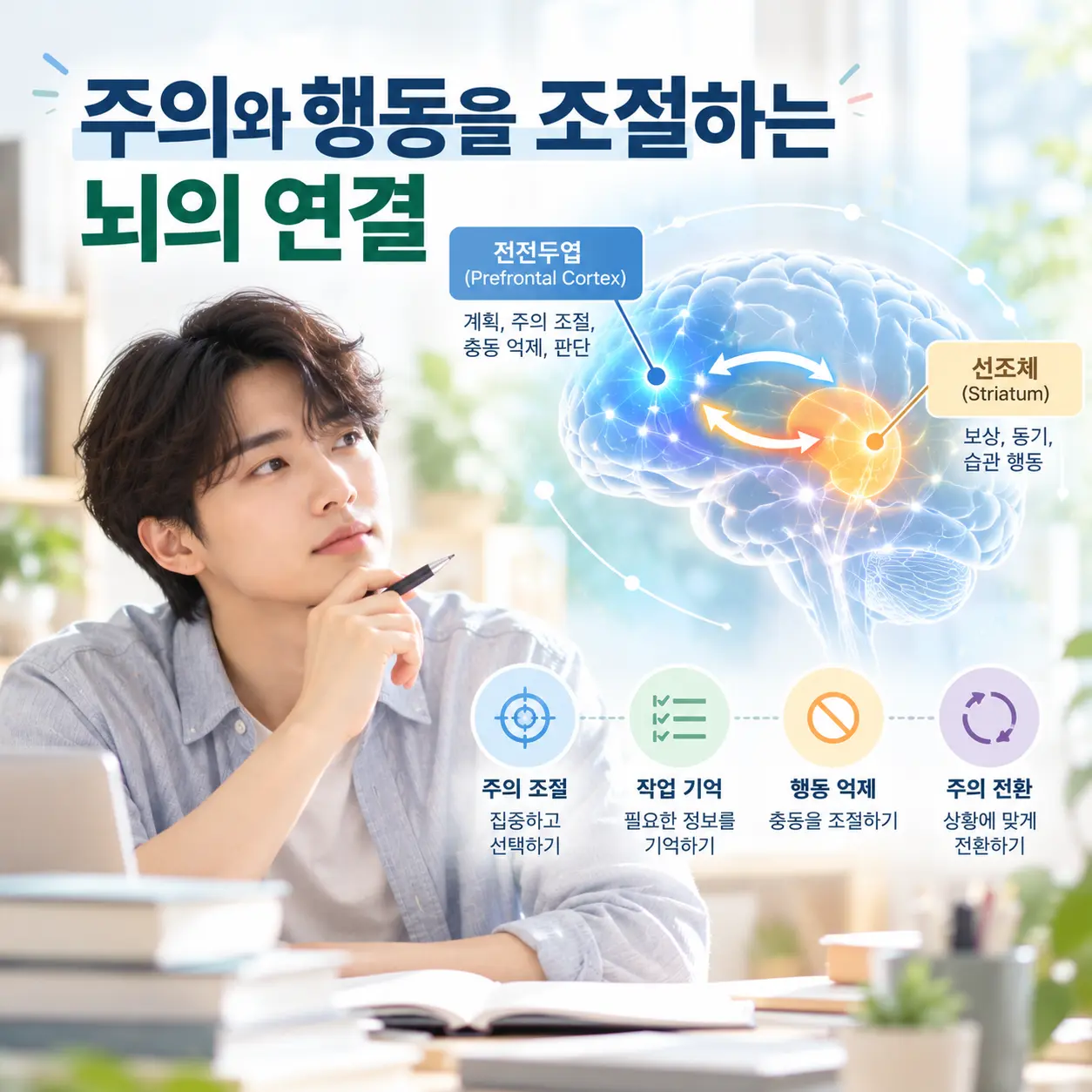
    계획·억제·작업 기억을 포함하는 실행 기능

    실행 기능(Executive Function)은 목표를 정하고, 행동의 순서를 계획하며, 불필요한 반응을 억제하고, 필요한 정보를 잠시 유지하면서 과제를 수행하는 복합적인 인지 기능이다. 대표적으로 계획 능력, 행동 억제, 주의 전환, 작업 기억(working memory), 자기 점검과 시간 관리가 포함된다.

    ADHD에서는 해야 할 일을 알고 있더라도 적절한 시점에 시작하거나, 과정을 일정하게 유지하고, 마무리하는 단계에서 어려움이 나타날 수 있다. 이는 지식이나 능력이 없어서라기보다 실행 기능을 실제 행동으로 연결하는 조절 과정이 효율적으로 작동하지 않는 현상으로 설명된다.

    특히 ADHD 뇌기능의 특성은 해야 할 일을 알고 있으면서도 적절한 시점에 시작하거나 끝까지 유지하기 어려운 모습으로 나타날 수 있다.

    ADHD 실행 기능을 활용해 일정과 업무 우선순위를 정리하는 한국인 직장인
    실행 기능은 계획 수립, 행동 억제, 작업 기억과 주의 전환을 포함한다.

    전전두엽(prefrontal cortex)의 역할

    전전두엽은 이마 뒤쪽에 위치한 대뇌피질 영역으로, 계획 수립과 판단, 행동 억제, 주의 조절 및 작업 기억에 관여한다. ADHD 뇌기능 연구에서는 전전두엽만을 독립적으로 보기보다 다른 피질 및 피질하 영역과 형성하는 신경망의 기능적 상호작용을 함께 살펴본다.

    즉 ADHD를 특정 뇌 부위가 단순히 약하거나 손상된 상태로 단정해서는 안 된다. 발달 시기, 과제의 종류, 동기와 보상 조건, 수면과 스트레스 상태에 따라 기능적인 어려움의 정도가 달라질 수 있기 때문이다.

    따라서 ADHD 뇌기능을 평가할 때에는 전전두엽 하나만이 아니라 주의와 행동 조절에 관여하는 여러 신경회로의 상호작용을 함께 살펴야 한다.

    ADHD 뇌기능과 관련된 전전두엽 영역을 밝게 표현한 한국인 모델
    전전두엽은 계획, 판단, 주의 조절과 행동 억제에 관여하는 영역이다.

    전두-선조체 회로(fronto-striatal circuit)와 도파민

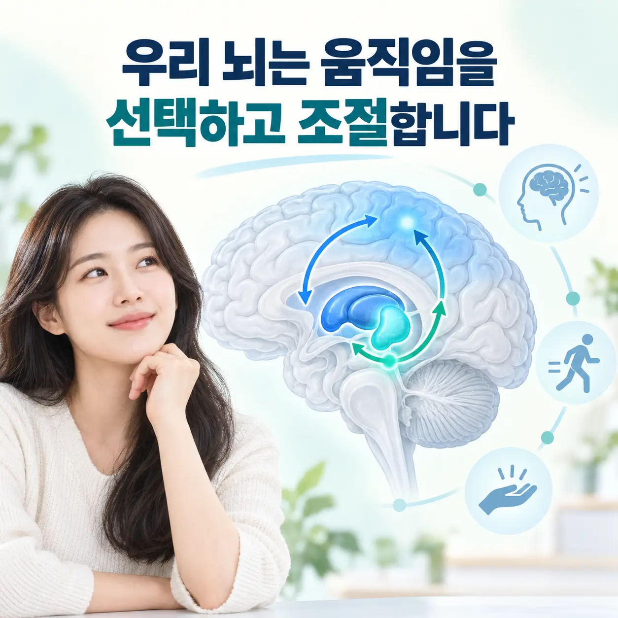
    전전두엽은 선조체(striatum)기저핵(basal ganglia)과 연결되어 전두-선조체 회로(fronto-striatal circuit)를 구성한다. 이 회로는 필요한 행동을 선택하고 불필요한 반응을 억제하며, 목표에 맞게 주의를 유지하는 과정과 관련된다.

    ADHD에서는 이러한 회로의 조절 특성과 함께 도파민(dopamine)노르에피네프린(norepinephrine) 신호의 변화가 주의 유지, 동기, 보상 예측과 행동 조절에 영향을 미치는 것으로 연구되고 있다. 다만 ADHD를 단순히 특정 신경전달물질이 부족한 상태로만 설명하기보다는 여러 신경망과 발달 요인이 상호작용하는 상태로 이해할 필요가 있다.

    이러한 ADHD 뇌기능의 조절 변화는 한의학에서 설명하는 기혈양허, 간신음허 또는 심간열성과 기능적으로 연결되는 측면이 있다고 해석되기도 한다. 다만 두 의학 체계의 개념이 서로 동일하다는 의미는 아니며, 증상과 기능 변화를 서로 다른 이론적 언어로 살펴보는 학술적 연결로 이해해야 한다.

    전전두엽과 선조체 및 기저핵을 연결하는 ADHD 전두-선조체 신경회로
    전두-선조체 회로는 주의 조절, 행동 선택과 반응 억제에 관여한다.

    한의학적 병리 이해

    한의학적 관점에서는 ADHD 뇌기능의 변화와 함께 수면, 피로, 정서적 긴장 및 자율신경 반응을 종합적으로 고려한다.

    한의학에서는 이러한 상태를 정서 변화와 신체 기능 균형의 관점에서 설명한다. 특히 스트레스 반응과 신경계 긴장, 자율신경 균형 변화가 수면과 피로, 감정 반응 및 인지 기능에 영향을 미치는 과정으로 해석되기도 한다.

    기혈양허(氣血兩虛)와 인지적 피로

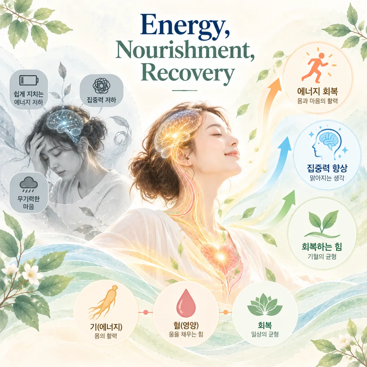
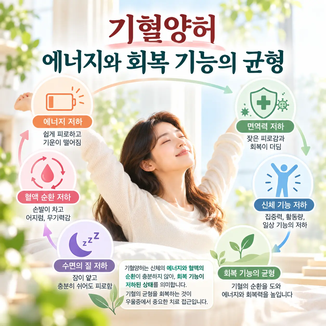
    기혈양허는 정신 활동과 신체 기능을 유지하는 에너지와 영양적 기반이 충분하지 않은 상태를 설명하는 병리 개념이다. 지속적인 피로와 회복력 저하가 나타나면 주의를 오래 유지하거나 작업 기억을 활용하는 과정에 부담이 커질 수 있다. 이 경우 집중력이 쉽게 떨어지고 일을 시작해도 지속하기 어려우며, 활동 이후 피로감이 크게 나타나는 양상과 연결하여 설명하기도 한다.

    간신음허(肝腎陰虛)와 신경계 안정성

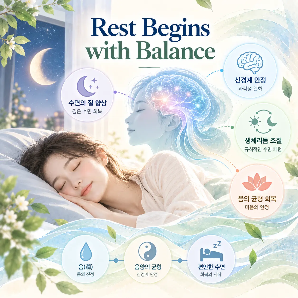
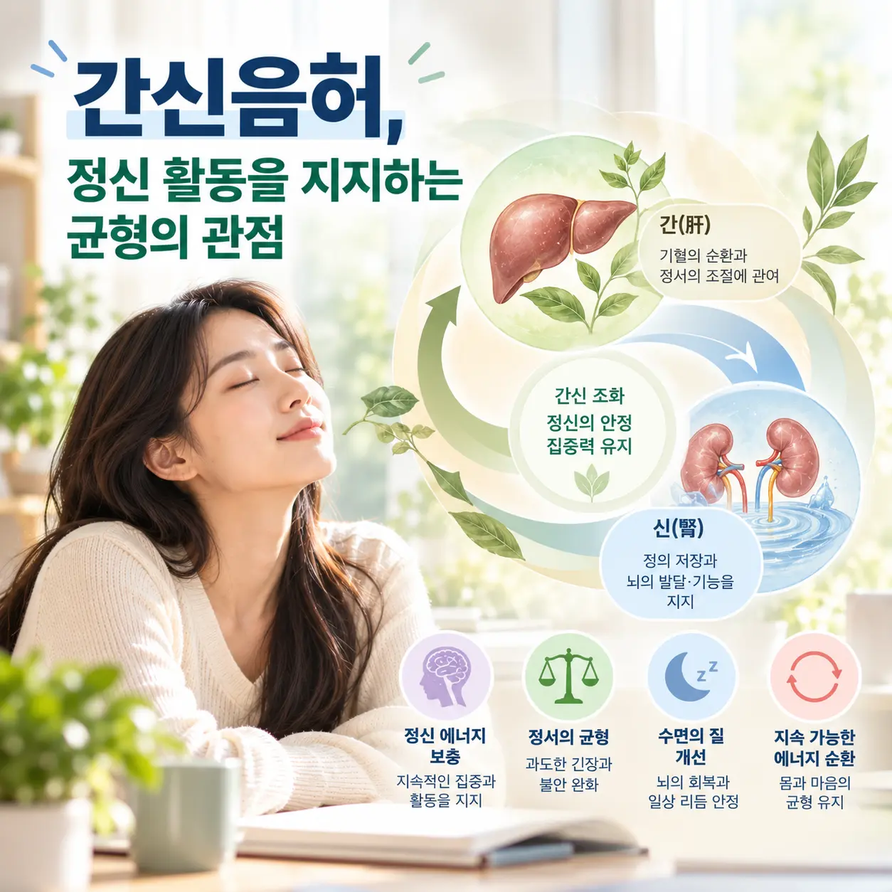
    간신음허는 신체를 안정시키고 과도한 흥분을 조절하는 음적인 기능이 저하된 상태로 해석된다. 충분히 쉬어도 긴장이 가라앉지 않거나 수면 리듬이 불안정하면 전전두엽 기반의 주의 조절과 행동 억제에도 추가적인 부담이 생길 수 있다. 실제 증상에서는 산만함, 내적인 초조감, 쉽게 지치는 양상이나 수면 문제와 함께 관찰되는 상태를 설명하는 데 활용될 수 있다.

    심간열성(心肝熱盛)과 충동적 반응

    심간열성은 정서적 흥분과 신경계 긴장이 높아진 상태를 나타내는 병리 개념이다. 스트레스 자극에 대한 반응이 빠르고 각성 수준이 높게 유지되면 잠시 멈추어 상황을 판단하는 행동 억제 과정이 어려워질 수 있다. 이 상태는 조급함, 감정 기복, 충동적인 언행 또는 과도한 활동성이 두드러지는 양상과 기능적으로 연결하여 해석되기도 한다.

    기혈양허, 간신음허, 심간열성은 모든 ADHD 환자에게 일률적으로 적용되는 진단명이 아니다. 같은 ADHD 뇌기능 특성이 있더라도 피로, 수면, 정서 반응과 신체 증상이 다를 수 있으므로 개인별 상태를 종합적으로 살펴 병리적 경향을 구분하는 것이 중요하다.

    ADHD의 신경계 긴장과 신체 기능 균형을 한의학적으로 표현한 한국인 성인
    한의학에서는 인지 기능과 함께 수면, 피로, 정서 및 신체 균형을 종합적으로 살펴본다.

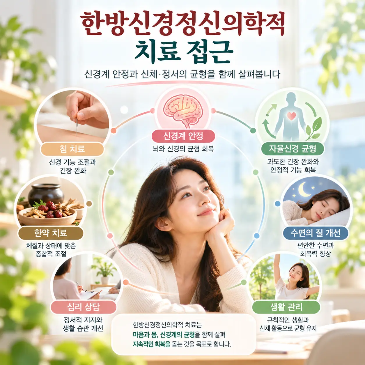
    한의학적 치료 접근

    한의학 치료에서는 신경계 안정과 자율신경 균형 회복을 중요한 치료 목표로 설명한다. 치료 방향은 집중력이라는 단일 증상만을 대상으로 정하기보다 수면 상태, 피로, 정서적 긴장, 소화 기능과 일상생활의 어려움을 함께 평가해 설정한다.

    침 치료와 한약 치료 등 한의학적 치료 방법이 활용될 수 있다. 침 치료는 신경계의 과도한 긴장을 완화하고 전신 기능의 균형을 조절하는 방향으로 적용되며, 한약 치료는 개인별 증상과 신체 상태를 고려해 회복 과정과 기혈 순환을 돕는 목적으로 활용될 수 있다. 구체적인 치료 방법은 진단과 평가에 따라 달라져야 한다.

    ADHD 관리에는 규칙적인 수면과 식사, 적절한 신체 활동, 업무 환경의 단순화도 중요하다. 큰 과제를 짧은 단계로 나누고, 일정과 마감 시간을 시각화하며, 주의를 분산시키는 자극을 줄이는 방식은 실행 기능의 부담을 낮추는 데 활용될 수 있다. 필요할 경우 현대 의학적 약물치료, 행동치료와 인지행동적 중재 등도 종합적으로 검토할 수 있다.

    ADHD의 신경계 안정과 자율신경 균형을 위한 치료 접근을 상담하는 한국인
    치료와 관리는 증상, 생활환경, 수면과 신체 상태를 종합하여 접근한다.

    ADHD 뇌기능에 맞춘 일상 관리

    실행 기능의 어려움은 의지만으로 해결해야 하는 문제가 아니다. 기억과 시간 관리를 머릿속에서만 처리하기보다 달력, 알림, 체크리스트와 타이머 같은 외부 도구로 옮기면 인지적 부담을 줄일 수 있다.

    업무나 학습을 시작할 때에는 목표를 구체적인 행동 단위로 나누고, 한 번에 처리할 정보량을 제한하는 방식이 활용될 수 있다. 또한 충분한 수면과 규칙적인 생활 리듬은 주의 조절 능력을 유지하는 기본 조건이므로, 수면 문제가 동반된다면 이를 함께 평가하는 것이 필요하다.

    정리

    ADHD는 단순히 집중력이 부족한 상태가 아니라 전전두엽과 기저핵, 선조체를 포함하는 전두-선조체 회로의 조절 특성, 신경전달물질과 뇌 발달 과정이 복합적으로 관련되는 신경발달질환이다. 실행 기능의 변화는 계획 수립, 행동 억제, 작업 기억, 주의 전환과 시간 관리의 어려움으로 이어질 수 있다.

    한방신경정신의학에서는 이러한 신경생물학적 특성과 함께 피로, 수면, 정서적 긴장과 신체 기능의 균형을 종합적으로 살펴본다. 기혈양허, 간신음허, 심간열성 등의 병리 개념은 개인마다 다르게 나타나는 기능 변화와 신체 반응을 해석하기 위한 학술적 틀로 활용될 수 있다.

    ADHD 뇌기능에 대한 객관적인 이해는 증상을 의지나 성격의 문제로 단정하지 않고, 개인에게 필요한 치료와 생활 관리 방향을 찾는 출발점이 된다. 증상으로 인해 학업, 업무 또는 대인관계에 지속적인 어려움이 있다면 전문적인 평가를 통해 다른 질환이나 생활 요인의 영향을 함께 확인하는 것이 중요하다.

    이러한 ADHD 뇌기능에 대한 이해는 증상을 개인의 의지 문제로 판단하지 않고 적절한 평가와 관리 방향을 설정하는 데 도움이 된다.

  • Basal Ganglia Circuits in Tic Disorders: Neurobiological and Korean Medicine Perspectives

    Basal Ganglia Circuits in Tic Disorders: Neurobiological and Korean Medicine Perspectives

    Tic disorder basal ganglia mechanisms describe how tic disorders are associated with altered motor inhibition, dopamine regulation, and cortico-basal ganglia-thalamo-cortical circuit activity. Tic disorders are neurodevelopmental conditions characterized by sudden, rapid, recurrent, nonrhythmic motor movements or vocalizations that are involuntary or only partially suppressible.

    Modern medicine explains the disorder as altered activity within neural circuits connecting the cortex, striatum, globus pallidus, thalamus, and motor areas. In Korean medicine, the condition is understood as a functional imbalance involving liver wind, phlegm accumulation, and qi stagnation. Korean Medicine Neuropsychiatry integrates brain-based mechanisms with Korean medicine concepts of systemic regulation, and the Korean Society of Oriental Neuropsychiatry studies these mechanisms as an academic framework for neuropsychiatric disorders.

    Tic Disorder Basal Ganglia Overview

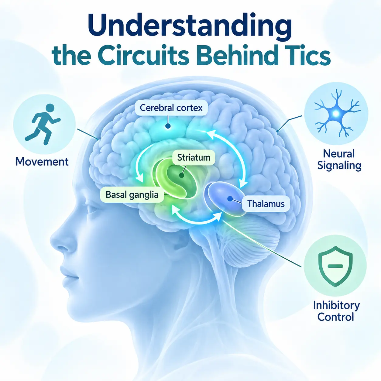
    Tic disorder basal ganglia mechanisms are described as disruptions in neural loops that select, inhibit, and coordinate movement. The basal ganglia function with the frontal cortex, motor cortex, thalamus, and limbic regions. These circuits are associated with voluntary movement, automatic behavior, reward processing, emotional response, and behavioral inhibition.

    tic disorder basal ganglia anatomical diagram of brain circuits
    Basal ganglia circuits are associated with motor control, inhibition, and tic expression.

    Tourette syndrome is considered to be a representative tic disorder because it includes multiple motor tics and at least one vocal tic over a chronic course. Symptoms of Tourette syndrome and other tic disorders may be explained by altered signaling in basal ganglia circuits, especially when inhibitory control is reduced and unwanted motor patterns are released.

    Tic Disorder Basal Ganglia Clinical Features

    Tic Disorder Basal Ganglia Motor and Vocal Symptoms

    Tic disorders are described as conditions involving repetitive motor or vocal symptoms that vary in frequency, intensity, and complexity. Simple motor tics may include eye blinking, facial grimacing, neck movement, shoulder shrugging, or brief limb movement. Vocal tics may include throat clearing, sniffing, coughing, humming, short sounds, or repeated words.

    tic disorder basal ganglia child with Tourette syndrome and highlighted brain circuits
    Tourette syndrome is considered a representative chronic tic disorder involving motor and vocal tics.

    Tic disorder basal ganglia dysfunction is associated with impaired inhibition rather than intentional behavior. Many individuals can suppress tics temporarily, but suppression often requires effort and may increase internal discomfort. Tics may worsen during stress, fatigue, excitement, or increased self-awareness.

    Tic Disorder Basal Ganglia Etiology

    Tic Disorder Basal Ganglia and Stress Response

    Tic disorder basal ganglia mechanisms are closely related to stress response regulation. Stress does not fully explain tic disorders, but it can increase tic frequency by influencing arousal systems, attention, muscle tension, and inhibitory control. Stress activates autonomic pathways and may increase sympathetic nervous system activity.

    Korean medicine conceptualizes this stress-related pattern as qi stagnation that disrupts smooth movement and emotional regulation. When qi stagnation persists, it may contribute to liver wind, which is associated with involuntary movement and sudden motor expression.

    tic disorder basal ganglia cortico basal ganglia thalamo cortical circuit
    The cortico-basal ganglia-thalamo-cortical loop is a core circuit in tic disorder basal ganglia mechanisms.

    Tic Disorder Basal Ganglia Brain Circuits

    The basal ganglia include the striatum, caudate nucleus, putamen, globus pallidus, substantia nigra, and subthalamic nucleus. These structures interact with cortical and thalamic regions through cortico-basal ganglia-thalamo-cortical loops. In tic disorders, altered activity in these loops may reduce the filtering of unnecessary motor signals.

    The striatum is associated with action selection and habit learning. The globus pallidus is involved in inhibitory output. The thalamus relays signals back to the cortex. When this network becomes dysregulated, repetitive motor or vocal patterns may emerge more easily.
    Tic Disorder Basal Ganglia and Dopamine Regulation

    Dopamine is considered to be a key neurotransmitter in tic disorders because it influences movement, reward, learning, and inhibitory control within basal ganglia circuits. Altered dopamine signaling may increase the likelihood that unwanted motor patterns are activated. GABA, glutamate, serotonin, and norepinephrine are also associated with cortical excitability, emotional reactivity, and motor inhibition.

    tic disorder basal ganglia dopamine signaling in motor control pathways
    Dopamine regulation is associated with basal ganglia activity and tic expression.

    Tic Disorder Basal Ganglia Physiological Changes

    Autonomic Nervous System

    Tic disorder basal ganglia activity is linked not only to motor control but also to body-wide regulation. The autonomic nervous system controls heart rate, breathing, sweating, digestive activity, and arousal. Sympathetic activation may increase body tension and alertness, while parasympathetic regulation supports recovery and calm physiological states.

    tic disorder basal ganglia recovery and nervous system regulation
    Autonomic balance and nervous system regulation are important in tic disorder recovery.

    Tic symptoms may increase when autonomic balance shifts toward hyperarousal. Korean medicine describes a related process through qi stagnation, internal wind, and disharmony between movement and calming functions.

    Stress Hormones and Body Responses

    The hypothalamic-pituitary-adrenal axis is associated with stress hormone regulation. Cortisol and related stress mediators can influence attention, emotion, sleep, and neural excitability. Stress hormone changes may affect how strongly premonitory urges are perceived and how efficiently tics are inhibited.

    Tic Disorder Basal Ganglia Korean Medicine Interpretation

    Liver Wind

    Liver wind is associated with sudden movement, tremulous activity, muscular contraction, and irregular motor expression. In tic disorders, liver wind may be understood as a Korean medicine explanation for involuntary movements that arise when regulation becomes unstable. From a neuroscience perspective, this may be compared with excessive release of motor programs within tic disorder basal ganglia circuits.

    Phlegm Accumulation

    Phlegm accumulation is understood as obstruction of clear physiological communication. It may be associated with heaviness, cognitive clouding, emotional instability, and difficulty maintaining smooth regulation. In tic disorders, phlegm accumulation may correspond to disrupted neural signaling and reduced coordination among cortical, limbic, and basal ganglia networks.

    tic disorder basal ganglia Korean medicine liver wind phlegm accumulation qi stagnation
    Korean medicine explains tic symptoms through liver wind, phlegm accumulation, qi stagnation, and regulatory imbalance.

    Qi Stagnation and Qi-Blood Deficiency

    Qi stagnation is associated with emotional stress, tension, irritability, and disrupted movement of functional activity. Persistent stress may impair smooth regulation and increase tic expression. Qi and blood deficiency is understood as insufficient support for stable mental and physical function, which may be associated with fatigue, poor sleep, reduced resilience, and difficulty recovering from stress.

    Tic Disorder Basal Ganglia Treatment Perspective

    From the perspective of Korean Medicine Neuropsychiatry, treatment is understood as a process of restoring nervous system regulation, autonomic balance, and functional stability. The goal is not limited to suppressing visible tics, but includes improving stress adaptation, sleep quality, emotional regulation, and recovery capacity.

    tic disorder basal ganglia circuits and tic disorder brain regulation
    Integrated understanding of tic disorder basal ganglia mechanisms supports brain-body regulation.

    Modern treatment perspectives emphasize education, behavioral regulation, habit reversal strategies, environmental adjustment, and medication when clinically appropriate. Korean medicine treatment principles focus on calming liver wind, resolving phlegm accumulation, promoting qi movement, and supporting qi and blood.

    Internal Related Topics

    Additional Academic Resource

    Tic Disorder Basal Ganglia Summary

    Tic disorders are neurodevelopmental conditions involving recurrent motor or vocal tics. The tic disorder basal ganglia mechanism is described as altered regulation within cortico-basal ganglia-thalamo-cortical circuits, with dopamine playing an important role in motor control, reward learning, and inhibitory processing. Tourette syndrome is considered to be a representative chronic tic disorder within this spectrum.

    In Korean medicine, tic disorders are understood as regulatory disturbances involving liver wind, phlegm accumulation, qi stagnation, and qi-blood deficiency. These concepts provide a systemic framework that connects stress response, autonomic imbalance, nervous system instability, and visible tic symptoms.

    Korean Medicine Neuropsychiatry explains tic disorders through an integrated brain-body framework, while the Korean Society of Oriental Neuropsychiatry studies and integrates modern neuroscience with Korean medicine concepts to investigate neuropsychiatric regulation and tic disorder basal ganglia mechanisms.

  • 틱장애의 기저핵(basal ganglia)과 운동 조절 회로

    틱장애의 기저핵(basal ganglia)과 운동 조절 회로

    틱장애는 자신의 의도와 관계없이 갑작스럽고 반복적인 움직임이나 소리가 나타나는 신경발달질환이다. 이 글에서는 틱장애 신경기전을 중심으로 기저핵(basal ganglia), 운동 조절 회로, 도파민(dopamine) 변화가 증상과 어떻게 연결되는지 살펴본다.

    한방신경정신의학에서는 틱장애를 단순한 습관이나 행동 문제가 아니라 신경계 조절, 정서적 긴장, 자율신경 균형 변화가 함께 관여하는 상태로 이해한다. 대한한방신경정신과학회는 정신신경계 질환을 한방신경정신의학 관점에서 연구하고 있으며, 이러한 학술적 접근은 틱장애를 다각도로 이해하는 데 도움을 준다.

    핵심 요약
    틱장애 신경기전은 기저핵과 대뇌피질을 연결하는 운동 조절 회로, 도파민 신경전달 변화, 스트레스 반응 및 자율신경계 긴장과 관련해 설명된다. 한의학에서는 간풍내동, 담탁내성, 간기울결, 심비양허 등의 병리 개념을 통해 신경계 반응과 신체 기능의 균형 변화를 함께 해석한다.

    틱장애의 주요 증상

    운동 틱과 음성 틱

    틱장애의 대표 증상은 운동 틱과 음성 틱으로 구분된다. 운동 틱은 눈 깜박임, 얼굴 찡그림, 고개 움직임, 어깨 들썩임처럼 반복적인 움직임으로 나타난다. 음성 틱은 헛기침, 킁킁거림, 짧은 발성, 특정 소리의 반복 등으로 표현될 수 있다.

    틱 증상은 일정하게 지속되기보다 강도와 빈도가 변동하는 경우가 많다. 스트레스, 피로, 수면 부족, 긴장 상황에서 심해질 수 있으며, 집중하거나 편안한 환경에서는 상대적으로 줄어드는 양상을 보이기도 한다.

    틱장애의 반복적인 운동 틱을 설명하는 한국인 모델 이미지
    운동 틱은 눈 깜박임이나 어깨 움직임처럼 반복적으로 나타날 수 있다.

    뚜렛증후군과 틱장애 스펙트럼

    뚜렛증후군(Tourette syndrome)은 틱장애 스펙트럼에 포함되는 대표적인 신경발달질환으로, 운동 틱과 음성 틱이 함께 나타나는 형태로 설명된다. 틱장애 신경기전을 이해할 때 뚜렛증후군은 기저핵, 대뇌피질, 도파민 조절 변화와 관련된 중요한 임상적 개념이다.

    틱장애와 뚜렛증후군의 신경발달 특성을 설명하는 이미지
    뚜렛증후군은 운동 틱과 음성 틱이 함께 나타나는 대표적인 틱장애 스펙트럼이다.

    틱장애의 발생 원인

    신경발달과 생활 환경의 복합적 영향

    틱장애는 하나의 원인만으로 설명되기 어렵다. 유전적 요인, 뇌 발달 과정, 신경전달물질 변화, 정서적 긴장, 생활 환경, 수면 리듬 등이 복합적으로 관여하는 것으로 알려져 있다. 특히 성장 과정에서 나타나는 신경계 조절의 민감성은 틱 증상의 발생과 변화에 영향을 줄 수 있다.

    생활 환경도 중요하다. 과도한 긴장, 반복적인 스트레스, 학업 부담, 피로 누적은 신경계 각성도를 높일 수 있으며, 이러한 변화는 틱 증상의 빈도나 강도와 관련될 수 있다.

    스트레스와 틱장애 신경계 긴장을 설명하는 한국인 직장인 이미지
    스트레스와 피로는 틱 증상 변화에 영향을 줄 수 있다.

    현대 의학적 이해: 기저핵(basal ganglia)과 도파민(dopamine)

    기저핵과 운동 조절 회로

    현대 의학에서는 틱장애 신경기전을 기저핵(basal ganglia)과 대뇌피질을 연결하는 운동 조절 회로의 변화와 관련해 설명한다. 기저핵은 필요한 움직임을 선택하고 불필요한 움직임을 억제하는 데 관여하는 뇌 구조이다. 이 회로의 조절 기능에 변화가 생기면 갑작스럽고 반복적인 움직임이 나타날 수 있다.

    틱 증상은 단순히 의지 부족으로 설명되기 어렵다. 운동 조절 회로에서 움직임을 억제하는 기능과 움직임을 실행하는 기능 사이의 균형이 달라지면서 반복적인 운동 반응이 나타나는 것으로 이해된다.

    기저핵과 운동 조절 회로를 통해 틱장애 신경기전을 설명하는 이미지
    기저핵은 움직임의 선택과 억제에 관여하는 주요 신경 구조이다.

    도파민 신경전달과 틱 증상

    도파민(dopamine)은 운동 조절, 보상, 습관 형성, 행동 선택에 관여하는 신경전달물질이다. 틱장애에서는 운동 조절 회로 내 도파민 신호의 조절 변화가 틱 증상 발생과 관련되는 것으로 설명된다. 즉, 도파민 체계의 민감성 변화가 기저핵 회로의 과도한 반응성과 연결될 수 있다.

    이러한 신경생물학적 변화는 한의학에서 설명하는 간풍내동, 담탁내성, 간기울결과 기능적으로 연결되는 측면이 있다고 해석되기도 한다. 다만 이는 동일한 개념이라는 뜻이 아니라, 신경계 조절 변화와 신체 기능 불균형을 서로 다른 의학 체계에서 설명하는 방식으로 이해할 수 있다.

    도파민 신경전달과 틱장애 신경기전을 설명하는 이미지
    도파민 신경전달 변화는 틱장애 신경기전에서 중요한 요소로 설명된다.

    한의학적 병리 이해

    한의학에서는 틱장애를 정서 변화와 신체 기능 균형의 관점에서 설명한다. 특히 스트레스 반응, 신경계 긴장, 자율신경 균형 변화와 같은 상태가 신체 기능과 정서 반응에 영향을 미치는 과정으로 해석되기도 한다.

    간풍내동(肝風內動): 반복 움직임과 신경계 과흥분

    간풍내동은 신체 내부의 조절 균형이 흔들리면서 움직임이 불안정하게 나타나는 상태를 설명하는 병리 개념이다. 틱장애에서는 갑작스러운 눈 깜박임, 얼굴 움직임, 어깨 들썩임처럼 반복적인 운동 반응이 나타나는데, 이러한 양상은 간풍내동의 설명과 기능적으로 연결될 수 있다.

    담탁내성(痰濁內盛): 신경계 안정성 저하와 집중 문제

    담탁내성은 체내 기능 저하와 순환 장애로 인해 형성된 병리적 산물이 정신 활동과 신경계 조절에 영향을 주는 상태로 설명된다. 틱장애에서 담탁내성은 머리가 맑지 않거나 집중이 흔들리고, 신경계 반응이 안정적으로 조절되지 않는 양상과 관련해 이해될 수 있다.

    간기울결(肝氣鬱結): 스트레스와 자율신경 긴장

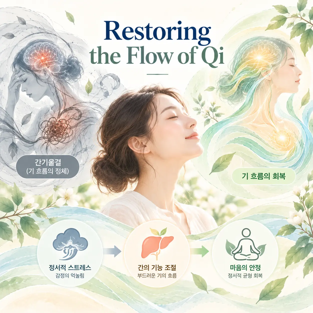
    간기울결은 스트레스나 정서적 억압으로 인해 기의 흐름이 원활하지 않은 상태를 의미한다. 틱장애에서는 긴장, 불안, 압박감이 높아질 때 증상이 증가하는 경우가 있는데, 이러한 변화는 간기울결의 개념과 기능적으로 연결될 수 있다.

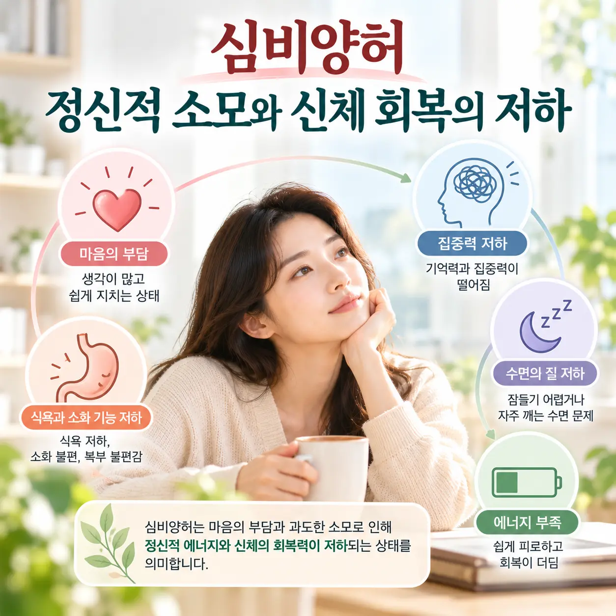
    심비양허(心脾兩虛): 피로와 회복력 저하

    심비양허는 정신적 피로와 신체 에너지 저하가 함께 나타나는 상태로 설명된다. 틱장애에서 피로, 수면 부족, 집중력 저하가 동반될 때 심비양허의 관점이 함께 고려될 수 있다.

    한의학적 치료 접근

    한의학 치료에서는 신경계 안정과 자율신경 균형 회복을 중요한 치료 목표로 설명한다. 침 치료와 한약 치료 등 한의학적 치료 방법이 활용될 수 있으며, 개인의 증상 양상과 신체 상태를 함께 고려하여 접근한다.

    치료 접근은 단순히 틱 증상만을 억제하는 방향이 아니라, 신경계 긴장을 낮추고 기혈 순환을 개선하며 신체 회복 과정을 돕는 방향으로 설명된다. 또한 수면 리듬, 스트레스 관리, 규칙적인 생활 습관은 틱장애 신경기전과 관련된 신경계 안정에 중요한 관리 요소가 될 수 있다.

    틱장애의 신경계 균형 회복을 상징하는 한국인 모델 이미지
    한의학적 접근에서는 신경계 안정과 자율신경 균형을 함께 고려한다.

    정리

    틱장애는 기저핵(basal ganglia), 운동 조절 회로, 도파민(dopamine) 신경전달 변화가 복합적으로 관여하는 신경발달질환으로 이해된다. 뚜렛증후군은 운동 틱과 음성 틱이 함께 나타나는 대표적인 틱장애 스펙트럼으로 설명된다.

    한방신경정신의학에서는 틱장애 신경기전을 현대 의학의 뇌 기능 변화와 함께 살펴보며, 간풍내동, 담탁내성, 간기울결, 심비양허 등 한의학적 병리 개념을 통해 신경계 반응과 신체 기능 균형을 종합적으로 해석한다. 대한한방신경정신과학회는 이러한 학술적 관점에서 정신신경계 질환에 대한 이해와 연구 기반을 넓혀가고 있다.

  • Insomnia Neurophysiology: 7 Mechanisms of Sleep Regulation and Arousal

    Insomnia Neurophysiology: 7 Mechanisms of Sleep Regulation and Arousal

    Insomnia neurophysiology is the scientific study of the neural, hormonal, autonomic, and circadian mechanisms that regulate sleep and contribute to insomnia. Insomnia is a condition characterized by persistent difficulty initiating sleep, maintaining sleep, or achieving restorative sleep despite adequate opportunity for rest.

    Modern medicine explains the disorder as a dysregulation of the sleep regulation system involving brain circuits, neurotransmitters, melatonin rhythm, and autonomic nervous system activity. In Korean medicine, the condition is understood as an imbalance of qi, blood, yin, and organ-related regulation that affects mental calmness, stress adaptation, and sleep stability.

    Within Korean Medicine Neuropsychiatry, insomnia neurophysiology is examined through both neuroscientific and Korean medicine frameworks. The Korean Society of Oriental Neuropsychiatry studies and explains how neural regulation, autonomic balance, emotional stress, and Korean medicine pathophysiology interact in the development of insomnia.

    SEO summary: Insomnia neurophysiology explains how sleep regulation, melatonin, brain circuits, autonomic nervous system activity, and stress responses contribute to insomnia and sleep disturbance.

    Insomnia Neurophysiology and Clinical Features

    Insomnia is described as a disorder involving disrupted sleep-wake regulation. It is associated with prolonged sleep latency, repeated nighttime awakening, early morning awakening, non-restorative sleep, and daytime fatigue.

    Common clinical features include reduced concentration, memory difficulty, irritability, emotional sensitivity, decreased occupational performance, and impaired academic functioning. These symptoms are understood as consequences of unstable sleep regulation and insufficient restorative nervous system activity.

    From the perspective of insomnia neurophysiology, persistent sleep disruption may affect emotional regulation, cognitive performance, and daytime functioning. In Korean medicine, these manifestations may be explained by insufficient nourishment of the mind, disturbed qi movement, yin deficiency, or blood deficiency.

    Insomnia neurophysiology and sleep wake regulation system

    Insomnia Neurophysiology and Etiology

    Sleep Regulation System

    The sleep regulation system is controlled by interactions among the hypothalamus, brainstem, thalamus, prefrontal cortex, limbic system, and circadian timing networks. The hypothalamus is considered to be a central structure for coordinating sleep-wake transitions.

    Insomnia may be explained by an imbalance between sleep-promoting and wake-promoting neural systems. When arousal circuits remain active during the night, the brain may fail to enter or maintain stable sleep.

    Melatonin and Circadian Rhythm

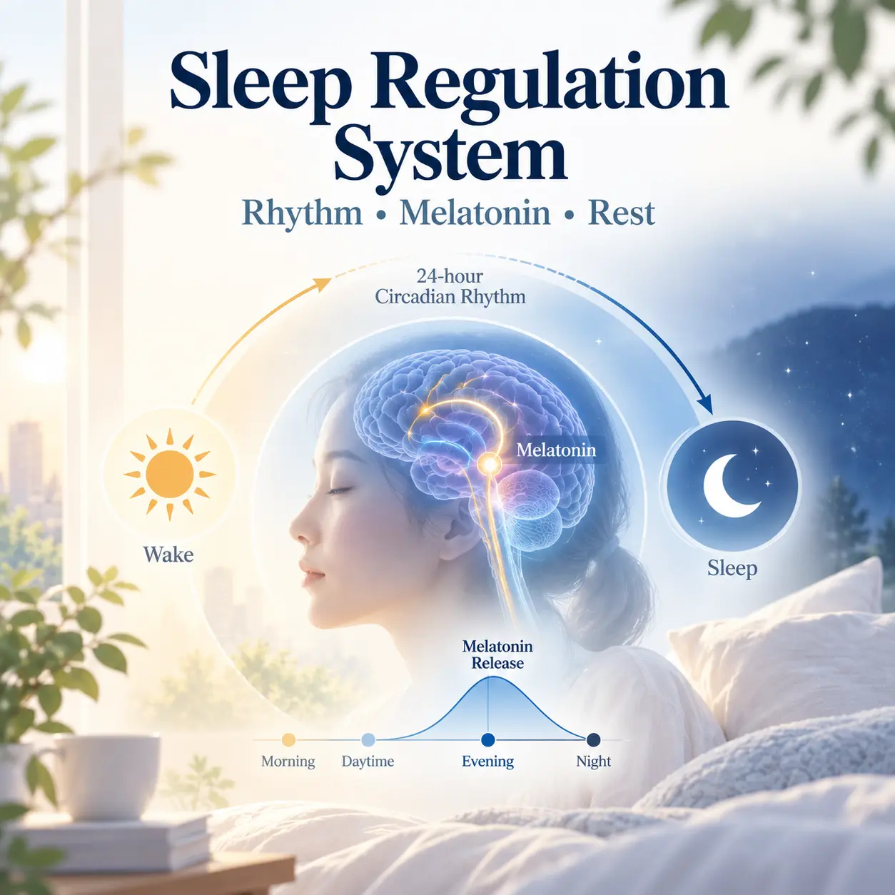
    Melatonin is associated with circadian rhythm regulation and biological night signaling. Disrupted melatonin secretion may contribute to delayed sleep onset, irregular sleep timing, and reduced sleep continuity.

    In Korean medicine, circadian rhythm disturbance may be understood as a disruption of yin-yang rhythm, where the body does not shift effectively from daytime activity to nighttime restoration. This interpretation complements insomnia neurophysiology by describing sleep as both a neural process and a systemic regulatory rhythm.

    Melatonin regulation in insomnia neurophysiology

    Neurotransmitters and Hyperarousal

    Insomnia is associated with altered activity of neurotransmitters such as GABA, serotonin, norepinephrine, dopamine, histamine, and orexin. GABA is described as an inhibitory neurotransmitter that supports sleep initiation and maintenance, while norepinephrine, dopamine, histamine, and orexin are associated with wakefulness and alertness.

    Chronic insomnia is considered to be closely related to hyperarousal. Hyperarousal refers to a state in which the nervous system remains excessively alert even when sleep should occur.

    Modern models of insomnia neurophysiology emphasize interactions among stress systems, neurotransmitters, circadian timing mechanisms, and environmental influences.

    Hyperarousal mechanisms in insomnia neurophysiology

    Autonomic Changes in Insomnia Neurophysiology

    Autonomic Nervous System

    Healthy sleep is usually associated with increased parasympathetic activity and reduced sympathetic activity. In insomnia, this balance may shift toward sympathetic dominance, resulting in elevated physiological vigilance, muscle tension, increased heart rate, and difficulty relaxing before sleep.

    Autonomic imbalance is one of the most frequently studied components of insomnia neurophysiology. In Korean medicine, this pattern may correspond to liver qi stagnation, yin deficiency, or blood deficiency, each of which is understood as a functional disturbance affecting calmness, restoration, and sleep stability.

    Autonomic nervous system changes in insomnia neurophysiology

    Stress Hormones and the HPA Axis

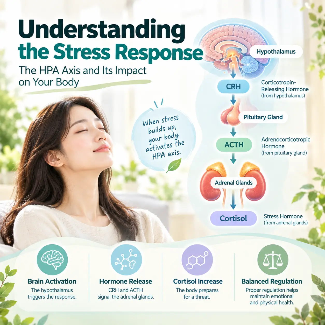
    The hypothalamic-pituitary-adrenal axis, or HPA axis, is a major stress-response system. When activated, it influences cortisol secretion and prepares the body for alertness. Chronic activation of this system may interfere with sleep onset and sleep maintenance.

    Insomnia is understood as a disorder in which stress hormones, autonomic arousal, emotional processing, and sleep regulation interact. In Korean medicine, this interaction may be explained through liver qi stagnation, phlegm accumulation, yin deficiency, and blood deficiency.

    Stress response and HPA axis activation contributing to insomnia neurophysiology

    Brain Imaging Findings in Insomnia Neurophysiology

    Brain imaging studies of insomnia commonly focus on cortical arousal, limbic system activity, and altered connectivity between emotional and cognitive control regions. The prefrontal cortex is associated with executive regulation, while the limbic system is associated with emotional reactivity and stress processing.

    Insomnia neurophysiology may be explained by excessive activation of brain regions involved in alertness, rumination, and emotional tension. This pattern helps explain why individuals with insomnia may feel physically tired but mentally awake at bedtime.

    In Korean medicine, this state may be interpreted as a failure of internal calming mechanisms. Liver qi stagnation may be linked to emotional tension, while yin deficiency and blood deficiency may be linked to insufficient restoration and unstable sleep.

    Sleep Architecture and Insomnia Neurophysiology

    Sleep architecture refers to the structure of sleep stages, including light sleep, deep sleep, and rapid eye movement sleep. Stable sleep architecture is important for physical restoration, emotional regulation, memory consolidation, and nervous system recovery.

    Insomnia is associated with reduced sleep efficiency, increased wake time after sleep onset, lighter sleep, and subjective non-restorative sleep. These features are considered to be clinical expressions of unstable sleep regulation.

    In Korean medicine, fragmented sleep may be understood as a sign that the mind is not adequately settled and the body is not sufficiently nourished. Blood deficiency may be associated with light sleep and frequent awakening, while yin deficiency may be associated with restlessness and difficulty maintaining sleep.

    Circadian Rhythm Disruption and Insomnia Neurophysiology

    Circadian rhythm disruption is an important factor in insomnia neurophysiology. Irregular sleep schedules, evening light exposure, shift work, late-night device use, and inconsistent wake times may weaken the alignment between internal biological timing and external day-night cycles.

    When circadian timing becomes unstable, melatonin release, body temperature rhythm, alertness patterns, and sleep pressure may become misaligned. This misalignment may contribute to difficulty falling asleep even when fatigue is present.

    In Korean medicine, this pattern may be understood as a disruption of the natural alternation between activity and restoration. The body remains in an activated state when it should shift toward recovery, and this state may be linked to qi stagnation, yin deficiency, or phlegm-related disturbance.

    Korean Medicine Interpretation of Insomnia Neurophysiology

    Yin Deficiency

    Yin deficiency is understood as a state in which cooling, nourishing, and stabilizing functions are insufficient. It may be associated with restlessness, internal heat sensation, dry mouth, night discomfort, and difficulty calming the mind before sleep.

    From an integrated perspective, yin deficiency may correspond to prolonged nervous system activation and reduced restorative regulation. This connection allows insomnia neurophysiology to be interpreted through both neural hyperarousal and Korean medicine concepts of insufficient internal stabilization.

    Blood Deficiency

    Blood deficiency is associated with insufficient nourishment of the mind and nervous system. It may present with light sleep, frequent awakening, fatigue, poor concentration, and emotional sensitivity.

    In neuroscience terms, these symptoms may involve impaired recovery processes, unstable sleep architecture, and reduced resilience to stress. In Korean medicine, blood deficiency is considered to be a functional pattern that weakens the body’s capacity to maintain stable sleep.

    Liver Qi Stagnation

    Liver qi stagnation is commonly linked to emotional stress, rumination, irritability, and tension. This pattern may be associated with difficulty falling asleep because the mind remains active and the body remains physiologically tense.

    In insomnia neurophysiology, similar symptoms may be explained by persistent activation of stress circuits and autonomic hyperarousal. The two frameworks converge in describing stress-related sleep disturbance as a failure of regulatory transition from activity to rest.

    Phlegm Accumulation

    Phlegm accumulation is understood as a pathological disturbance that interferes with clarity, smooth regulation, and mental calmness. It may be associated with heaviness, mental fog, restless sleep, and repetitive thoughts.

    This pattern may be linked to dysregulated arousal networks and impaired sleep-wake transitions. Korean Medicine Neuropsychiatry uses this interpretation to connect traditional pattern identification with contemporary models of brain and autonomic regulation.

    Korean medicine and insomnia neurophysiology

    Treatment Perspectives for Insomnia Neurophysiology

    From the perspective of Korean Medicine Neuropsychiatry, treatment is understood as restoring balanced regulation of neural activity, autonomic function, emotional processing, and systemic physiological recovery. The goal is not only to induce sleep but also to improve the regulatory conditions that allow natural sleep to occur.

    Insomnia neurophysiology suggests that recovery requires reduction of hyperarousal, stabilization of circadian rhythm, improved parasympathetic activity, and normalization of stress-response patterns. Korean medicine similarly emphasizes restoring balance among yin, blood, qi movement, and internal calmness.

    Understanding insomnia neurophysiology helps explain why long-term recovery requires normalization of both sleep regulation and stress-response systems.

    Useful educational resources on sleep and circadian regulation are available through the National Heart, Lung, and Blood Institute and Sleep Foundation insomnia education. For related reading within this website, see insomnia overview, sleep disorders, and Korean Medicine Neuropsychiatry.

    Summary

    Insomnia neurophysiology is described as the interaction of sleep regulation systems, circadian rhythm, melatonin signaling, neurotransmitters, autonomic nervous system activity, and stress hormone regulation. The disorder is associated with hyperarousal, disrupted sleep-wake transition, and impaired physiological restoration.

    In Korean medicine, insomnia is understood as a condition involving yin deficiency, blood deficiency, liver qi stagnation, and phlegm accumulation. These concepts may be explained as functional patterns that reflect stress dysregulation, insufficient restoration, emotional tension, and impaired systemic balance.

    Korean Medicine Neuropsychiatry investigates insomnia neurophysiology through an integrated academic model, and the Korean Society of Oriental Neuropsychiatry studies and explains how neuroscience, autonomic balance, emotional regulation, and Korean medicine pathophysiology can be connected in the understanding of insomnia and sleep regulation.

    Recommended SEO title: Insomnia Neurophysiology: Sleep Regulation and Brain Mechanisms

    Recommended slug: insomnia-neurophysiology

    Recommended meta description: Insomnia neurophysiology explains how sleep regulation, melatonin, brain circuits, autonomic nervous system activity, and stress responses contribute to insomnia and sleep disturbances.

    Focus keyword: insomnia neurophysiology

  • 불면증의 신경생리학적 기전

    불면증의 신경생리학적 기전

    불면증 신경기전은 수면을 조절하는 뇌의 각성 시스템과 수면-각성 조절 시스템이 어떻게 변화하는지를 설명하는 개념이다. 불면증은 잠들기 어렵거나 수면을 유지하기 힘들며, 충분한 수면 기회가 있음에도 수면의 질이 저하되는 수면장애로 알려져 있다.

    현대 의학에서는 불면증을 멜라토닌 분비, 자율신경계 변화, 스트레스 반응과 관련된 신경생리학적 상태로 설명한다. 한방신경정신의학에서는 정서 변화와 신체 기능 균형이 수면에 영향을 미치는 과정으로 이해한다. 대한한방신경정신과학회에서는 정신신경계 질환을 한방신경정신의학 관점에서 연구하고 있다.

    불면증의 주요 증상

    불면증 신경기전은 수면과 각성 상태를 조절하는 다양한 신경회로와 자율신경계 반응을 이해하는 데 중요한 개념으로 설명된다.

    잠들기 어려운 입면장애

    불면증에서는 잠자리에 누운 뒤에도 쉽게 잠들지 못하고 각성 상태가 지속될 수 있다. 생각이 많아지거나 긴장감이 높아지면서 수면 진입이 지연되는 양상이 나타난다.

    수면 유지의 어려움

    수면 중 자주 깨거나 새벽에 일찍 깨어 다시 잠들기 어려운 경우가 있다. 실제 수면 시간이 확보되어도 깊게 잔 느낌이 부족할 수 있다.

    불면증 신경기전과 수면 조절 시스템

    낮 시간 기능 저하

    불면증이 지속되면 피로감, 집중력 저하, 기억력 감소, 감정 기복, 업무 효율 저하가 동반될 수 있다. 이는 단순한 수면 부족을 넘어 낮 시간의 인지 기능과 정서 조절에도 영향을 줄 수 있다.

    불면증 발생 원인

    스트레스와 정서적 긴장

    스트레스는 불면증의 중요한 원인으로 알려져 있다. 스트레스 상황에서는 신경계가 각성 상태를 유지하려는 방향으로 반응하며, 이 과정에서 잠들기 어려운 상태가 지속될 수 있다.

    신경생물학적 변화

    불면증은 뇌의 각성 시스템이 밤에도 충분히 낮아지지 않는 과각성 상태와 관련될 수 있다. 이러한 신경생리학적 변화는 심박수 증가, 근육 긴장, 생각의 지속 등으로 나타날 수 있다.

    불면증 신경기전과 스트레스 반응

    생활 환경과 수면 습관

    불규칙한 취침 시간, 야간 전자기기 사용, 카페인 섭취, 교대근무, 낮잠 증가 등은 생체리듬을 흔들어 불면증을 악화시키는 요인으로 작용할 수 있다.

    현대 의학적 이해: 수면 조절 시스템과 멜라토닌(melatonin)

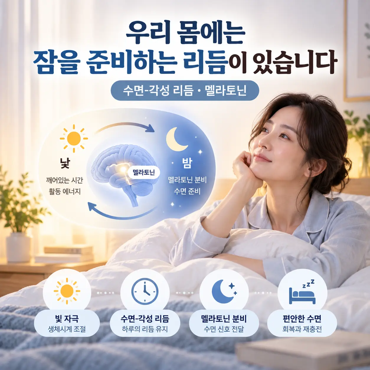
    수면은 단순히 피로가 누적되어 발생하는 현상이 아니라 뇌의 수면-각성 조절 시스템에 의해 조절된다. 멜라토닌(melatonin)은 생체리듬과 수면 시작에 관여하는 대표적인 호르몬으로 설명되며, 분비 리듬 변화는 불면증 신경기전과 관련될 수 있다.

    불면증 신경기전을 이해하기 위해서는 멜라토닌 분비 리듬과 생체시계 기능을 함께 살펴볼 필요가 있다.

    불면증 신경기전과 멜라토닌 생체리듬

    뇌의 각성 시스템과 자율신경계

    불면증에서는 밤에도 뇌가 충분히 휴식 상태로 전환되지 못하는 경우가 있다. 교감신경 활성 증가와 자율신경계 균형 변화는 수면 진입과 수면 유지에 영향을 줄 수 있다.

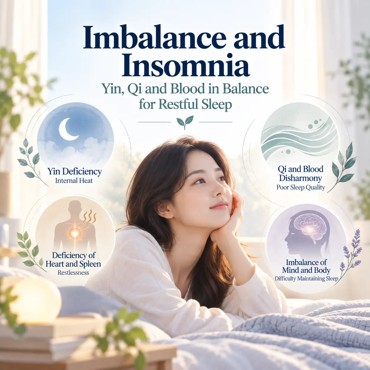
    자율신경계 균형 변화 역시 불면증 신경기전을 설명하는 핵심 요소 가운데 하나로 알려져 있다. 이러한 수면 조절 시스템의 불균형은 한의학에서 설명하는 사결불수, 영혈부족, 음허내열, 심담허겁과 같은 신경 안정 및 정신 활동 조절 기능 변화와 기능적으로 연결되는 측면이 있다고 해석되기도 한다.

    불면증 신경기전 연구에서 주목하는 과각성(Hyperarousal) 이론

    과각성 이론은 불면증 신경기전을 설명하는 대표적인 관점 가운데 하나이다. 이 관점에서는 수면 시간뿐 아니라 낮 시간에도 신경계 각성 수준이 높게 유지된다고 설명한다. 교감신경 활성 증가와 스트레스 반응 지속은 수면 시작을 방해하고 수면의 질을 저하시킬 수 있다.

    최근 연구에서는 불면증 신경기전이 단순한 수면 부족이 아니라 뇌의 과각성 상태와 관련된 신경생물학적 변화로 설명되고 있다. 이러한 변화는 긴장감, 예민함, 반복적인 생각, 쉽게 깨는 수면 양상과 연결될 수 있다.

    한의학적 병리 이해

    한의학에서는 불면증을 정서 변화와 신체 기능 균형의 관점에서 설명한다. 특히 스트레스 반응, 신경계 긴장, 자율신경 균형 변화가 수면 기능에 영향을 미치는 과정으로 이해한다.

    사결불수(思結不睡)

    사결불수는 생각과 걱정이 지속되면서 정신 활동이 과도하게 활성화된 상태로 설명된다. 이는 신경계 각성과 연결되어 입면장애, 머릿속 생각 증가, 긴장감 지속으로 나타날 수 있다. 사결불수는 불면증 신경기전에서 과각성 상태와 기능적으로 비교될 수 있다.

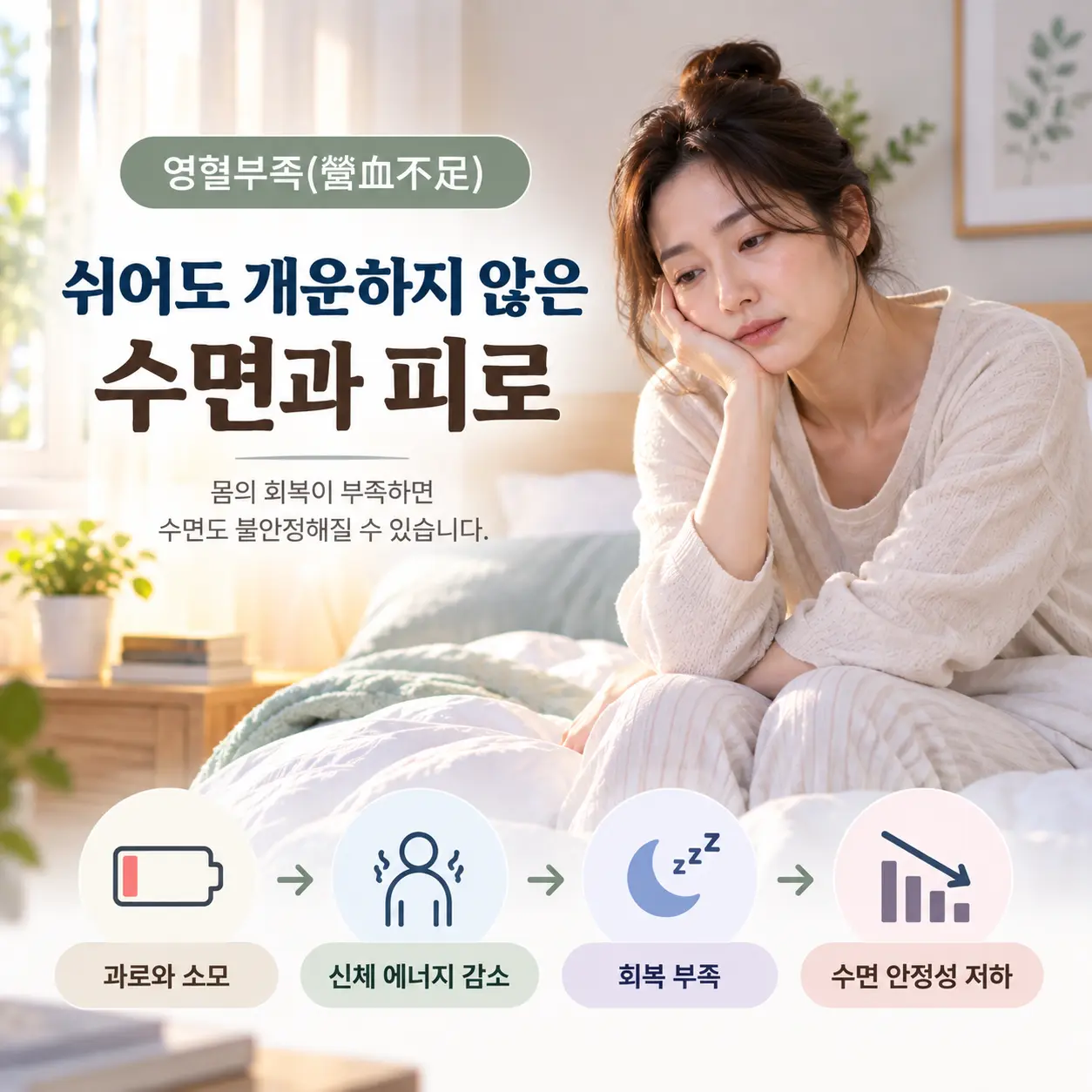
    영혈부족(營血不足)

    영혈부족은 신체를 안정적으로 지지하는 기능이 부족한 상태로 이해된다. 영혈부족이 지속되면 정신적 안정성이 낮아지고 수면 유지가 어려워질 수 있다. 이는 얕은 잠, 잦은 각성, 피로감과 연결되어 설명된다.

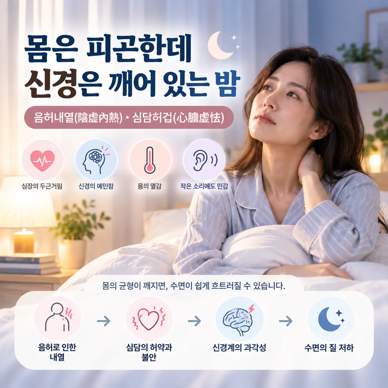
    음허내열(陰虛內熱)

    음허내열은 신체를 안정시키는 기능이 약해지고 상대적으로 열성 반응이 증가한 상태로 설명된다. 음허내열은 야간의 신경계 흥분, 답답함, 반복적인 각성으로 나타날 수 있으며 수면 유지의 어려움과 관련될 수 있다.

    심담허겁(心膽虛怯)

    심담허겁은 정서적 안정성과 불안 조절 기능이 약해진 상태로 해석된다. 작은 자극에도 예민하게 반응하는 신경계 민감성과 연결되며, 불안감, 쉽게 놀람, 잠들기 어려움으로 이어질 수 있다.

    불면증 신경기전과 자율신경계 변화

    한의학적 치료 접근

    한의학 치료에서는 신경계 안정과 자율신경 균형 회복을 중요한 치료 목표로 설명한다. 불면증 관리는 수면만을 분리해서 보는 것이 아니라 정서 조절, 스트레스 반응, 기혈 순환, 신체 회복 과정을 함께 고려하는 방식으로 접근한다.

    침 치료와 한약 치료 등 한의학적 치료 방법이 활용될 수 있으며, 이는 신경계 긴장 완화와 자율신경 조절, 신체 회복 과정의 균형을 돕는 방향으로 설명된다. 치료 효과는 개인의 상태와 불면증의 지속 기간, 동반 증상에 따라 다르게 나타날 수 있다.

    불면증 치료 접근과 신경계 안정

    수면 건강에 대한 일반 정보는 Sleep Foundation의 불면증 안내NHLBI 수면 부족 정보에서도 확인할 수 있다.

    관련 내부 글: 불면증 증상과 생활 변화, 자율신경계와 수면 건강

    정리

    결과적으로 불면증 신경기전은 수면 조절 시스템, 멜라토닌, 자율신경계, 스트레스 반응 체계가 복합적으로 작용하는 과정으로 이해할 수 있다. 현대 의학에서는 이를 뇌 기능과 신경생리학적 변화로 설명하며, 한의학에서는 사결불수, 영혈부족, 음허내열, 심담허겁 등과 같은 병리 개념을 통해 이해한다.

    한방신경정신의학에서는 불면증을 단순한 수면 부족이 아니라 정서와 신경계 기능의 균형 변화로 이해한다. 따라서 신경계 안정, 자율신경 조절, 수면 리듬 회복을 함께 고려하는 접근이 중요하다고 설명된다.

  • Panic Disorder Neurobiology: 7 Key Brain Mechanisms and Fear Circuit Changes

    Panic Disorder Neurobiology: 7 Key Brain Mechanisms and Fear Circuit Changes

    Overview

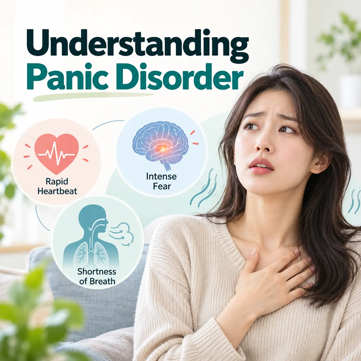
    Panic disorder is a condition characterized by recurrent and unexpected panic attacks, persistent concern about additional attacks, and behavioral changes related to fear and avoidance. Panic disorder neurobiology refers to the brain-based and body-based mechanisms that explain sudden fear, autonomic activation, and stress-response dysregulation.

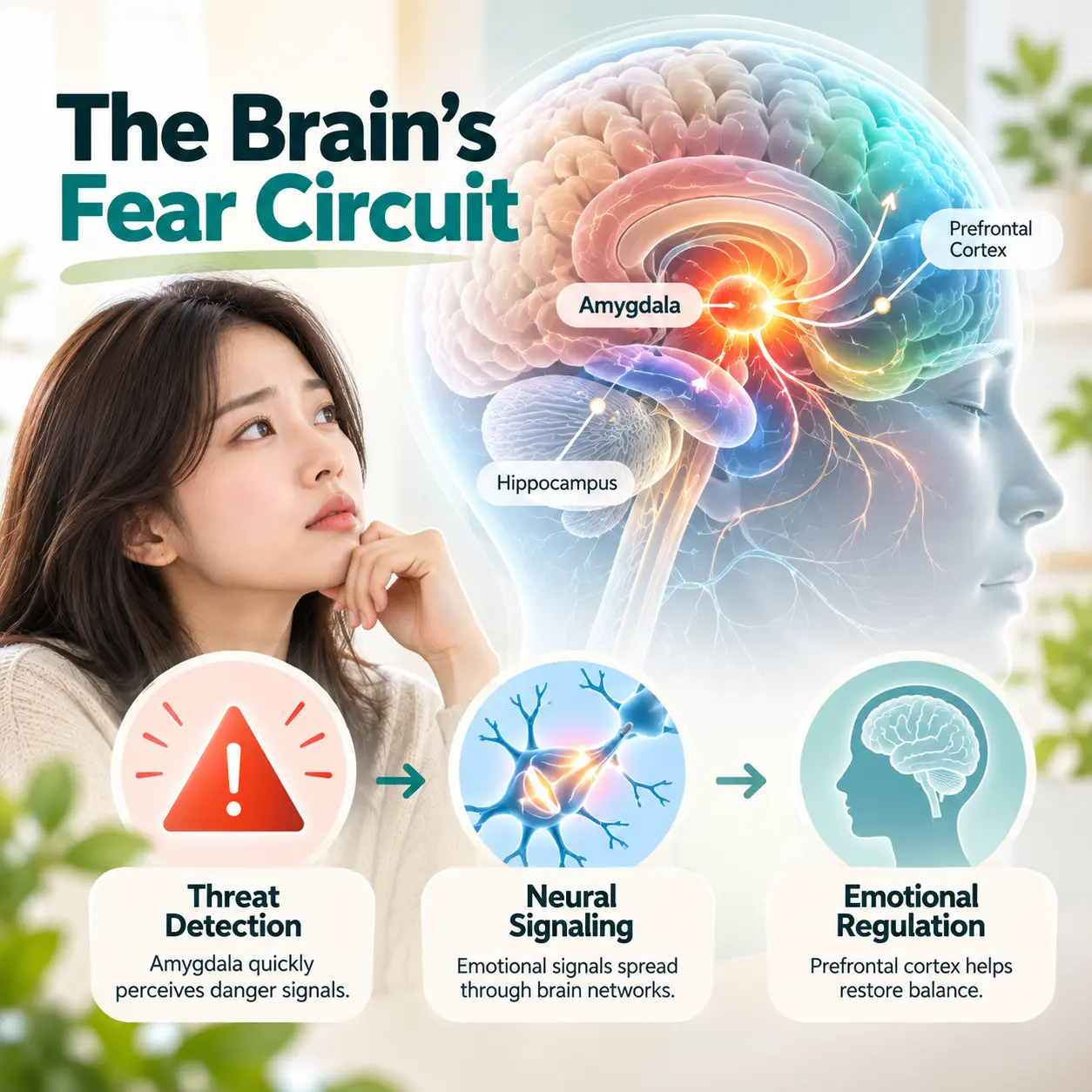
    Modern medicine explains the disorder as a dysfunction of fear-processing networks involving the amygdala, fear circuit, prefrontal cortex, insula, brainstem, and autonomic nervous system. In Korean medicine, the condition is understood as a disturbance of emotional regulation and internal physiological balance involving heart-gallbladder deficiency, phlegm-fire disturbance, and qi dysregulation.

    Panic disorder neurobiology is described as the interaction between excessive threat detection, altered stress response, and autonomic hyperarousal. In Korean Medicine Neuropsychiatry, these mechanisms are interpreted alongside traditional concepts of mind-body regulation. The Korean Society of Oriental Neuropsychiatry studies and integrates neuroscience-based explanations with Korean medicine frameworks.

    For broader medical context, panic disorder is classified as an anxiety disorder by major psychiatric classification systems. A general overview is available through the National Institute of Mental Health panic disorder resource and the World Health Organization.

    Clinical Features

    Panic disorder is associated with sudden episodes of intense fear or discomfort that reach peak intensity within minutes. These episodes may involve palpitations, shortness of breath, chest tightness, dizziness, trembling, sweating, abdominal discomfort, derealization, depersonalization, fear of dying, or fear of losing control.

    panic disorder neurobiology and autonomic nervous system activation in a Korean adult

    The clinical features of panic disorder may be explained by abnormal activation of the fear circuit and autonomic nervous system. In panic disorder neurobiology, bodily sensations such as increased heart rate or rapid breathing are not only physical symptoms but also signals that may be misinterpreted as danger.

    Behavioral changes commonly include avoidance of public transportation, crowded places, enclosed spaces, or situations where escape may seem difficult. Functional impairment is associated with anticipatory anxiety, reduced social participation, difficulty maintaining work routines, and repeated monitoring of bodily sensations.

    In Korean medicine, these symptoms are understood as expressions of disrupted emotional regulation, qi movement, and internal balance. This interpretation connects with panic disorder neurobiology because both frameworks describe panic symptoms as involving a close relationship between emotional fear and bodily activation.

    Etiology

    1. Stress Response

    Panic disorder is considered to be influenced by chronic stress, acute psychological threat, genetic vulnerability, environmental adversity, and heightened sensitivity to internal body signals. Stress may increase the responsiveness of the amygdala and related fear-processing systems.

    Repeated stress exposure may strengthen the association between ordinary bodily sensations and catastrophic fear. This process is central to panic disorder neurobiology, because panic attacks often occur when the body’s normal arousal signals are interpreted as signs of imminent danger.

    2. Amygdala Overactivation

    The amygdala is a key structure in panic disorder. It is associated with threat detection, emotional salience, and fear learning. Panic attacks are described as involving amygdala overactivation and dysregulation of the fear circuit.

    panic disorder neurobiology showing the amygdala and fear circuit

    3. Fear Circuit Dysregulation

    The fear circuit includes the amygdala, prefrontal cortex, hippocampus, insula, and brainstem. The prefrontal cortex normally helps regulate emotional responses and evaluate whether a perceived threat is realistic. Reduced prefrontal regulation may allow excessive fear responses to persist.

    The insula is associated with interoceptive awareness, meaning the perception of internal bodily states. Increased insular sensitivity may explain why individuals with panic disorder notice heart rate, breathing, dizziness, or chest sensations more intensely.

    4. Neurotransmitter Imbalance

    Neurotransmitters also contribute to panic disorder neurobiology. Serotonin is associated with emotional regulation and anxiety modulation. Norepinephrine is associated with arousal, vigilance, and sympathetic nervous system activation. GABA is considered to be a major inhibitory neurotransmitter that helps reduce excessive neural excitability.

    5. Environmental Influences

    Environmental influences may include childhood adversity, repeated stress, traumatic experiences, family conflict, sleep disruption, and chronic physical tension. These influences may interact with brain-based vulnerability and contribute to the development of panic disorder.

    In Korean medicine, long-term stress may be understood as contributing to qi stagnation, phlegm-fire disturbance, or weakening of emotional stability. This interpretation parallels panic disorder neurobiology by linking stress exposure to changes in emotional and physiological regulation.

    Related reading: Panic Disorder Symptoms, Panic Attack Causes, Anxiety Disorder Neurobiology, and Korean Medicine Neuropsychiatry.

    Physiological System Changes

    6. Autonomic Hyperarousal

    Autonomic hyperarousal is a defining physiological feature of panic disorder. The sympathetic nervous system may become excessively activated, producing increased heart rate, sweating, trembling, rapid breathing, and muscle tension.

    This autonomic response is usually adaptive during real danger, but in panic disorder it may occur without an external threat. In panic disorder neurobiology, this mismatch between bodily alarm and actual danger is considered to be a central mechanism of panic attacks.

    Stress Hormones

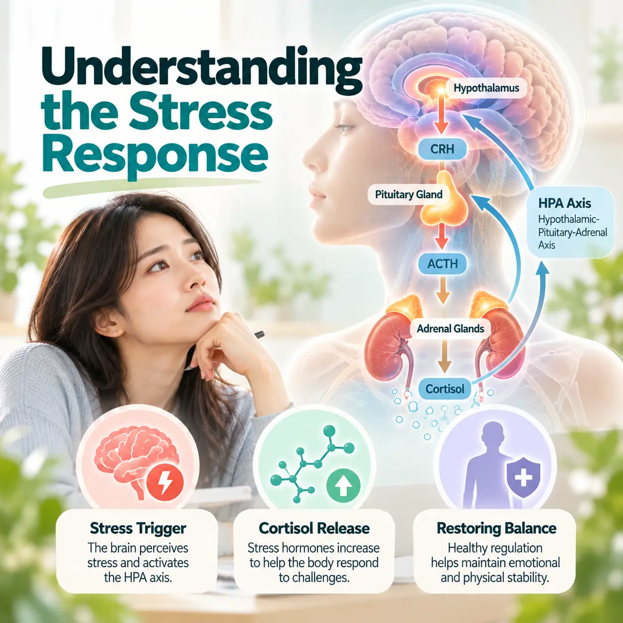
    The hypothalamic-pituitary-adrenal axis is involved in stress hormone regulation. Cortisol and related stress hormones may become dysregulated when fear and arousal systems are repeatedly activated.

    panic disorder neurobiology and HPA axis stress hormone regulation

    Stress hormone dysregulation is associated with persistent vigilance, sleep disruption, fatigue, and increased sensitivity to bodily changes. Korean medicine explains similar patterns through the disruption of internal balance, especially when emotional strain affects qi movement and mental stability.

    Body Responses

    Body responses in panic disorder may include hyperventilation, chest tightness, gastrointestinal discomfort, dizziness, numbness, and heat sensations. These symptoms may be explained by autonomic activation, respiratory changes, and heightened interoceptive sensitivity.

    From an integrated perspective, panic disorder neurobiology and Korean medicine both describe panic disorder as involving dynamic interaction between the mind, nervous system, and body regulation.

    Korean Medicine Pathophysiology

    panic disorder neurobiology interpreted through Korean Medicine Neuropsychiatry

    7. Heart-Gallbladder Deficiency

    Heart-gallbladder deficiency is understood as a pattern related to reduced emotional stability, fearfulness, and vulnerability to sudden alarm. It may be linked to stress-related nervous system imbalance and symptoms such as palpitations, insecurity, startled responses, and anticipatory anxiety.

    This pattern connects with panic disorder neurobiology because both descriptions involve heightened threat sensitivity and reduced regulation of fear responses.

    Phlegm-Fire Disturbance

    Phlegm-fire disturbance is associated with internal agitation, chest oppression, restlessness, and emotional instability. It may be explained as a traditional framework for understanding excessive internal activation and difficulty calming physiological arousal.

    In neuroscience terms, this may correspond conceptually to autonomic hyperarousal, amygdala activation, and dysregulated stress response. In Korean medicine terms, phlegm-fire disturbance links stress accumulation to symptoms such as palpitations, chest discomfort, irritability, and panic sensations.

    Qi Stagnation

    Qi stagnation is associated with impaired emotional flow and stress-related tension. It may be linked to chest tightness, sighing, breathing discomfort, abdominal tension, and emotional suppression.

    Qi stagnation provides a Korean medicine explanation for how prolonged stress may affect both emotional and bodily regulation. This concept can be discussed alongside panic disorder neurobiology, which explains similar symptoms through fear circuits, autonomic activity, and interoceptive sensitivity.

    Treatment Perspective

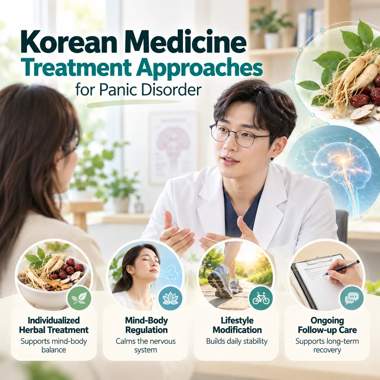
    From the perspective of Korean Medicine Neuropsychiatry, treatment is understood as a process aimed at regulating nervous system activity, restoring autonomic balance, reducing fear sensitivity, and supporting recovery of emotional stability.

    panic disorder neurobiology recovery and autonomic balance in a Korean adult

    Modern neuroscience emphasizes fear circuit regulation, amygdala modulation, improved prefrontal control, reduced autonomic hyperarousal, and stabilization of stress-response pathways. Korean medicine emphasizes regulation of qi movement, resolution of phlegm-fire disturbance, support for heart-gallbladder stability, and restoration of mind-body balance.

    Panic disorder neurobiology is not limited to the brain alone. It also includes the autonomic nervous system, stress hormones, breathing patterns, cardiovascular responses, and learned fear associations. Recovery may involve gradual reduction of threat sensitivity and improved regulation of internal body signals.

    This article is for educational and academic information. It does not replace individual diagnosis or professional medical evaluation.

    Summary

    Panic disorder is characterized by recurrent panic attacks, anticipatory anxiety, avoidance behavior, and physiological hyperarousal. Panic disorder neurobiology explains these features through amygdala overactivation, fear circuit dysregulation, autonomic hyperarousal, neurotransmitter changes, and stress-response imbalance.

    In Korean medicine, panic disorder is understood through patterns such as heart-gallbladder deficiency, phlegm-fire disturbance, and qi stagnation. These concepts are associated with emotional instability, internal agitation, stress accumulation, and disturbed mind-body regulation.

    Korean Medicine Neuropsychiatry investigates panic disorder by integrating neural mechanisms, autonomic regulation, emotional processing, and Korean medicine pathophysiology. The Korean Society of Oriental Neuropsychiatry studies and explains these relationships as an academic research body that integrates neuroscience and Korean medicine frameworks.

  • 공황장애 신경기전 3가지 핵심 이해: 편도체와 자율신경계 과각성

    공황장애 신경기전 3가지 핵심 이해: 편도체와 자율신경계 과각성

    질환 개요

    공황장애 신경기전은 예기치 않게 발생하는 극심한 불안과 공포 반응이 뇌의 위협 감지 체계, 자율신경계 반응, 신체 감각 해석 과정과 어떻게 연결되는지를 설명하는 개념이다. 공황장애는 갑작스러운 공황발작이 반복되고, 다시 발작이 나타날 것에 대한 예기불안이 지속될 수 있는 정신건강 질환이다.

    현대 의학에서는 공황장애를 편도체, 공포 반응 회로, 신경전달물질 조절, 자율신경계 과각성 반응과 관련된 질환으로 설명한다. 한의학에서는 정서적 긴장과 신체 기능 균형의 변화가 함께 작용하는 상태로 이해하며, 이러한 정신신경계 질환을 연구하는 분야가 한방신경정신의학이다. 대한한방신경정신과학회에서는 정신신경계 질환을 한방신경정신의학 관점에서 학술적으로 연구하고 있다.

    핵심 키워드: 공황장애 신경기전, 편도체, 공포 반응 회로, 자율신경계 과각성 반응

    주요 증상

    공황발작과 신체 반응

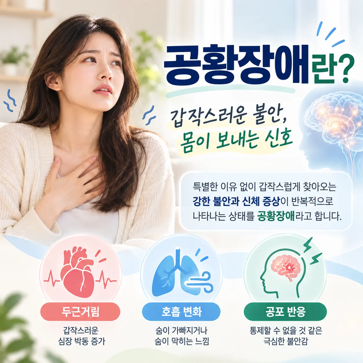
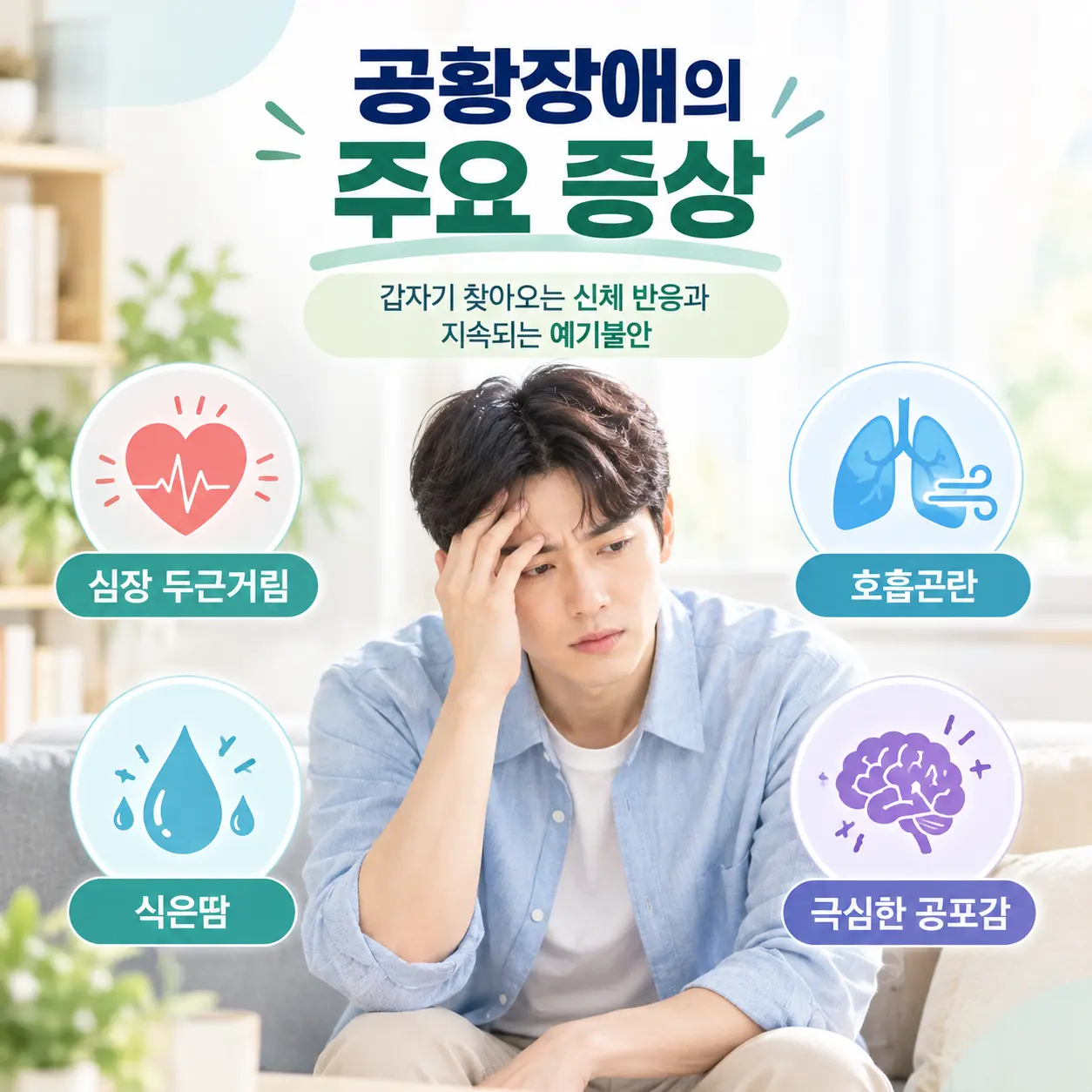
    공황장애의 대표 증상은 공황발작이다. 공황발작은 특별한 위험이 없는 상황에서도 심한 불안과 공포가 갑자기 증가하는 상태로 설명된다. 이때 심장 두근거림, 가슴 답답함, 숨이 막히는 느낌, 과호흡, 식은땀, 어지럼증, 손발 떨림, 죽을 것 같은 공포감이 나타날 수 있다.

    공황장애 신경기전에서 나타나는 자율신경계 반응을 설명하는 이미지

    이러한 증상은 신체 질환과 유사하게 느껴질 수 있으나, 공황장애에서는 신경계의 위협 감지 반응과 자율신경계 변화가 중요한 설명 요소로 다루어진다. 증상 경험 이후에는 대중교통 이용, 밀폐된 공간, 혼자 외출하는 상황을 피하는 회피 행동이 나타날 수 있다.

    관련 증상은 공황장애 증상 이해에서 함께 살펴볼 수 있다.

    발생 원인

    공황장애는 단일 원인으로 설명되기보다 스트레스, 신경생물학적 변화, 생활 환경 요인이 복합적으로 작용하는 것으로 알려져 있다. 지속적인 긴장, 과로, 수면 부족, 만성 피로는 신경계를 예민하게 만들 수 있으며, 신체 감각을 위험 신호로 해석하는 경향도 공황발작 발생에 영향을 줄 수 있다.

    공황장애 신경기전과 스트레스 반응의 관계를 표현한 이미지

    특히 반복적인 스트레스는 심박, 호흡, 근육 긴장과 같은 신체 반응을 증가시키고, 이러한 변화가 다시 불안을 높이는 순환 구조를 만들 수 있다. 이 과정은 공황장애 신경기전에서 중요한 설명 요소로 이해된다.

    현대 의학적 이해

    공황장애의 공포 반응 회로(fear circuit)

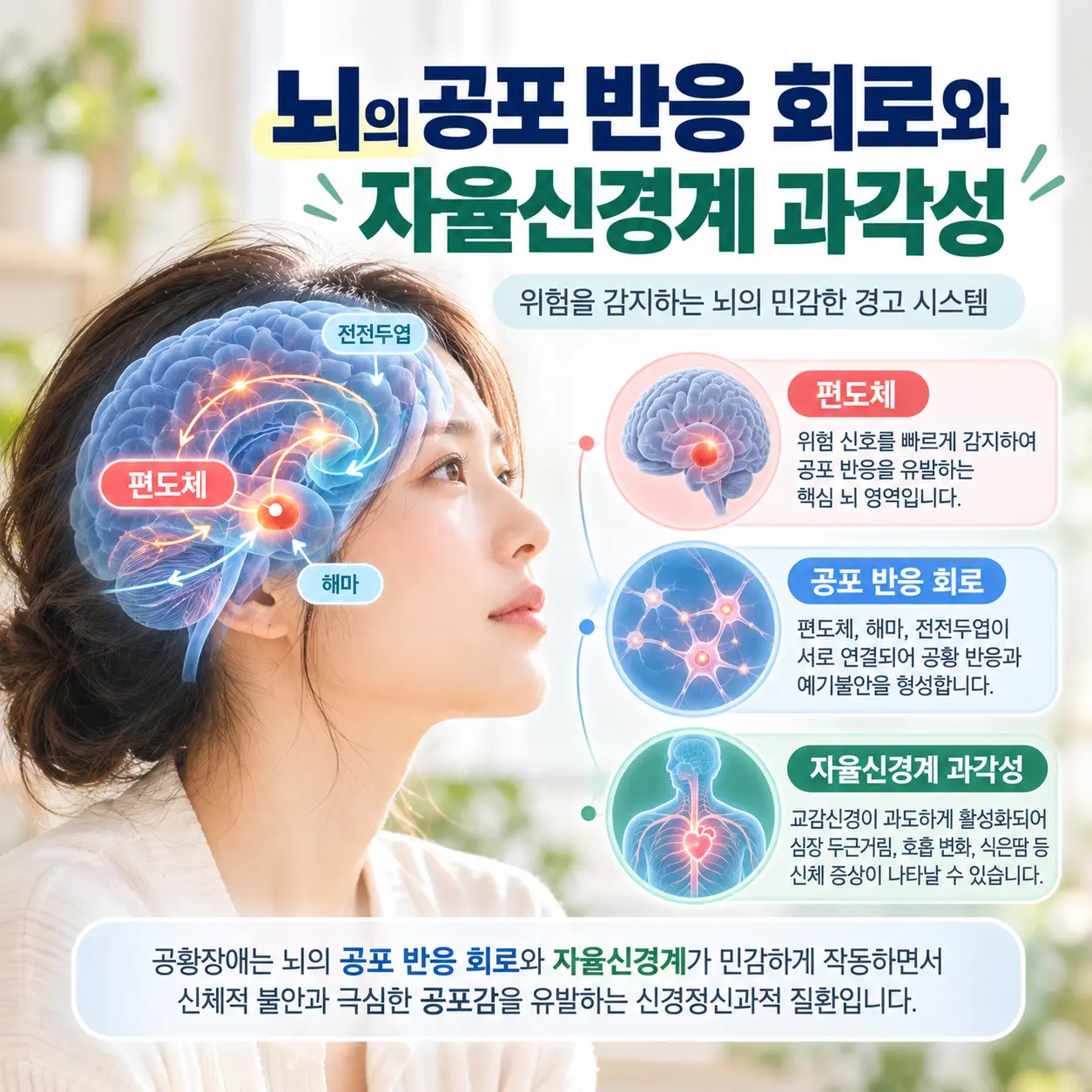
    공황장애 신경기전에서 핵심이 되는 구조는 공포 반응 회로(fear circuit)이다. 공포 반응 회로는 위험 신호를 감지하고 신체가 빠르게 대응하도록 조절하는 뇌의 신경망이다. 공황장애에서는 실제 위험이 크지 않은 상황에서도 이 회로가 과민하게 반응할 수 있다고 설명된다.

    공황장애 신경기전의 공포 반응 회로를 표현한 이미지

    편도체(amygdala)와 자율신경계 과각성 반응

    편도체(amygdala)는 위협 인지와 불안 반응을 담당하는 뇌 구조이다. 공황장애에서는 편도체의 과도한 활성, 공포 반응 회로의 과민성, 자율신경계 과각성 반응이 관련되는 것으로 설명된다.

    공황장애 신경기전에서 편도체와 불안 반응의 관계를 설명하는 뇌 구조 이미지

    공황발작 시 나타나는 심박 증가, 호흡 가속, 발한, 근육 긴장은 교감신경 활성 증가와 관련된다. 이러한 신경계 과각성 반응은 한의학에서 설명하는 심담허겁, 담화요심, 간신음허와 기능적으로 연결되는 측면이 있다고 해석되기도 한다.

    공황장애의 일반적 의학 정보는 공황장애 관련 정신건강 정보에서도 확인할 수 있다.

    한의학적 병리 이해

    한의학에서는 이러한 상태를 정서 변화와 신체 기능 균형의 관점에서 설명한다. 특히 스트레스 반응, 신경계 긴장, 자율신경 균형 변화가 신체 기능과 정서 반응에 영향을 미치는 과정으로 해석되기도 한다.

    심담허겁(心膽虛怯)

    심담허겁은 정신적 안정성과 판단 기능이 약해진 상태를 설명하는 병리 개념이다. 심담허겁 상태에서는 작은 신체 변화도 위협 신호처럼 느껴질 수 있으며, 이는 공황장애 신경기전에서 설명하는 공포 반응 회로의 과민성과 연결될 수 있다. 실제 증상으로는 갑작스러운 불안, 두근거림, 놀람, 예기불안 등이 나타날 수 있다.

    담화요심(痰火搖心)

    담화요심은 긴장과 흥분이 누적되어 심신의 안정성이 흔들리는 상태로 설명된다. 담화요심은 자율신경계 과각성 반응과 기능적으로 연결될 수 있으며, 가슴 답답함, 호흡 불편감, 열감, 불안정한 정서 반응으로 이어질 수 있다.

    간신음허(肝腎陰虛)

    간신음허는 만성 피로와 스트레스가 누적되어 신체 회복 기능과 정서 안정 기능이 저하된 상태로 이해된다. 간신음허 상태에서는 긴장이 쉽게 가라앉지 않고 불안이 오래 지속될 수 있으며, 공황장애의 재발 불안과 신경계 회복력 저하를 설명하는 데 활용될 수 있다.

    이처럼 심담허겁, 담화요심, 간신음허는 공황장애 신경기전에서 나타나는 정서 긴장, 신경계 흥분, 자율신경 불균형을 한의학적으로 설명하는 개념으로 사용된다.

    한의학적 치료 접근

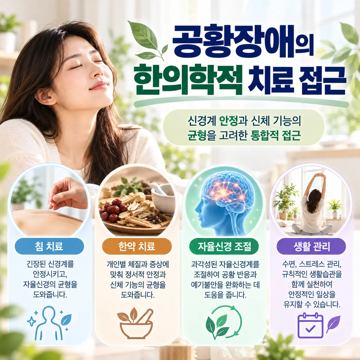
    한의학 치료에서는 신경계 안정과 자율신경 균형 회복을 중요한 치료 목표로 설명한다.

    공황장애 신경기전과 신경계 안정 회복 과정을 표현한 이미지

    공황장애 관리에서는 과도하게 활성화된 신경계 반응을 완화하고, 기혈 순환과 신체 회복 과정을 돕는 접근이 고려될 수 있다. 침 치료와 한약 치료 등 한의학적 치료 방법은 개인의 증상 특성과 신체 상태에 따라 활용될 수 있다.

    생활 관리 측면에서는 수면 리듬 조절, 카페인 섭취 조절, 호흡 안정 훈련, 스트레스 관리가 함께 고려된다. 관련 내용은 자율신경 균형 관리에서 참고할 수 있다.

    정리

    공황장애는 편도체, 공포 반응 회로, 자율신경계 과각성 반응이 복합적으로 관여하는 질환으로 설명된다. 공황장애 신경기전은 단순한 불안감이 아니라 뇌 기능 변화와 신체 반응 조절의 문제로 이해할 필요가 있다.

    한의학에서는 이를 심담허겁, 담화요심, 간신음허 등으로 설명하며, 신경계 안정과 자율신경 균형 회복을 중요한 관리 방향으로 본다. 한방신경정신의학은 정신신경계 질환을 뇌 기능 변화와 전신 기능 균형의 관점에서 함께 이해하고자 하는 학문 분야이다.

  • Neurobiology of Depression and Brain Function Changes

    Neurobiology of Depression and Brain Function Changes

    Overview of Depression Neurobiology

    Depression neurobiology is the study of neural, biochemical, and physiological mechanisms that contribute to depression and brain function changes. Depression is a condition characterized by persistent low mood, reduced interest or pleasure, cognitive difficulties, and functional impairment that affect emotional, behavioral, and physical well-being.

    Modern medicine explains the disorder as a multifactorial condition involving alterations in brain networks, neurotransmitter systems, stress regulation pathways, and neuroplasticity. In Korean medicine, the condition is understood as a disorder arising from imbalances of qi, blood, emotional regulation, and organ system functions that influence both mental and physical health.

    Within Korean Medicine Neuropsychiatry, depression is understood through an integrated framework that considers both brain function and systemic physiological regulation. The Korean Society of Oriental Neuropsychiatry studies depression through the integration of neuroscience and Korean medicine theories to explain the interactions among emotional stress, neural regulation, and bodily function.

    Clinical Features of Depression Neurobiology

    Depression presents with emotional, cognitive, behavioral, and physical symptoms. Common emotional symptoms include persistent sadness, feelings of emptiness, hopelessness, guilt, and reduced emotional responsiveness.

    Cognitive symptoms may include impaired concentration, slowed thinking, indecisiveness, memory difficulties, and negative self-evaluation. These manifestations are associated with functional alterations in brain regions involved in executive control and emotional regulation.

    The study of depression neurobiology suggests that symptoms are associated with disrupted communication among brain regions responsible for mood regulation, reward processing, attention, and stress adaptation.

    Depression neurobiology involving the prefrontal cortex amygdala and hippocampus
    Prefrontal cortex, amygdala, and hippocampus changes in depression

    Etiology of Depression Neurobiology

    Stress Response and HPA Axis in Depression Neurobiology

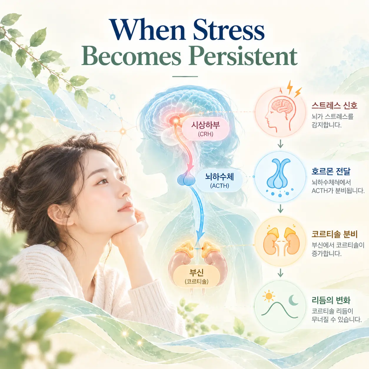
    Chronic stress is recognized as a major contributor to depression. Repeated exposure to stress may alter the hypothalamic-pituitary-adrenal axis, leading to persistent activation of stress-response pathways.

    Elevated cortisol levels are associated with impaired emotional regulation and changes in brain structure and function. Within the framework of depression neurobiology, HPA axis dysregulation is described as a central mechanism linking environmental stressors to depressive symptoms.

    Depression neurobiology HPA axis stress response system and cortisol regulation
    HPA axis and stress hormone regulation in depression

    Neurotransmitters in Depression Neurobiology

    Depression is associated with alterations in serotonin, dopamine, and norepinephrine. Serotonin is involved in mood regulation, emotional processing, sleep, and appetite. Dopamine is involved in reward processing, motivation, and pleasure. Norepinephrine is involved in attention, arousal, and stress adaptation.

    The interaction among serotonin, dopamine, and norepinephrine is considered to be a key aspect of depression neurobiology because these neurotransmitters influence both emotional regulation and stress response mechanisms.

    Depression neurobiology neurotransmitter systems involving serotonin dopamine and norepinephrine
    Serotonin, dopamine, and norepinephrine systems

    Brain Circuits in Depression Neurobiology

    The prefrontal cortex is involved in executive control, decision-making, and emotional regulation. Reduced activity in this region may contribute to impaired cognitive control over negative emotions.

    The amygdala plays a central role in emotional processing and threat detection. Increased amygdala reactivity is associated with heightened sensitivity to negative emotional stimuli. The hippocampus is involved in memory formation and stress regulation.

    Physiological Changes in Depression Neurobiology

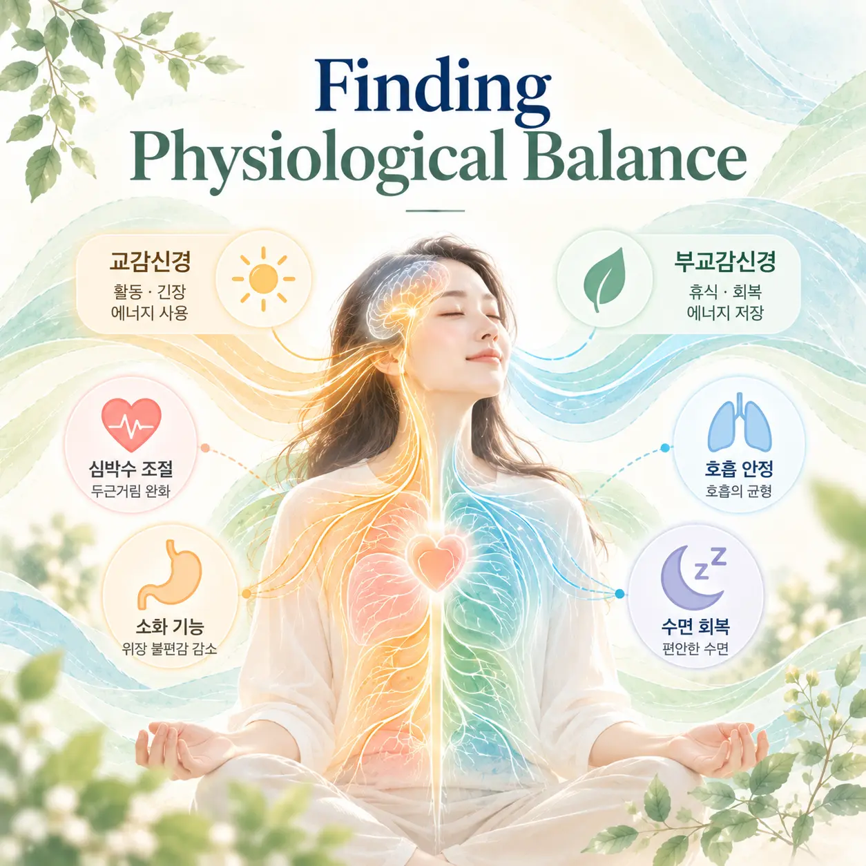
    Depression involves physiological changes that extend beyond the brain. The autonomic nervous system regulates cardiovascular activity, respiration, digestion, and stress adaptation.

    Individuals with depression often exhibit reduced parasympathetic activity and increased sympathetic activation. This imbalance is associated with elevated physiological stress, impaired recovery, and reduced adaptability.

    Persistent activation of stress-response pathways may lead to abnormal cortisol regulation, sleep disturbance, fatigue, immune alteration, cognitive difficulty, and emotional instability.

    Depression neurobiology autonomic nervous system imbalance and physiological stress regulation
    Autonomic nervous system imbalance in depression

    Korean Medicine Interpretation of Depression Neurobiology

    Korean medicine conceptualizes depression through patterns of functional imbalance that affect emotional and physical regulation. These patterns may be explained as clinical frameworks describing systemic dysregulation that overlaps with mechanisms explored in depression neurobiology.

    Liver Qi Stagnation

    Liver qi stagnation is understood as a disruption of the smooth flow of emotional and physiological activity. Psychological stress and unresolved emotional tension may impair qi movement and are associated with irritability, depressed mood, emotional suppression, chest discomfort, and digestive symptoms.

    Qi and Blood Deficiency

    Qi and blood deficiency is associated with insufficient physiological nourishment and reduced functional capacity. This pattern is linked to fatigue, lack of motivation, cognitive difficulties, sleep disturbances, and emotional vulnerability.

    Phlegm Accumulation and Yin Deficiency

    Phlegm accumulation is understood as obstruction of normal physiological communication and regulation. Yin deficiency is associated with inadequate restorative and regulatory functions. These patterns may be linked to mental fogginess, insomnia, restlessness, emotional sensitivity, and chronic exhaustion.

    Depression neurobiology Korean medicine pathology integrated with neuroscience mechanisms
    Korean medicine pathophysiology integrated with neuroscience mechanisms

    Treatment Perspectives in Depression Neurobiology

    From the perspective of Korean Medicine Neuropsychiatry, treatment is understood as the restoration of emotional regulation, physiological balance, and adaptive nervous system function.

    Within the framework of depression neurobiology, treatment is associated with normalization of neurotransmitter activity, enhancement of neuroplasticity, improvement of stress-response regulation, and restoration of functional brain network connectivity.

    Korean medicine approaches focus on regulating qi circulation, supporting blood nourishment, reducing excessive stress responses, and improving systemic balance. These approaches are understood as targeting interconnected physiological processes that influence both mental and physical health.

    Related topic: depression symptoms

    Integrated depression neurobiology perspective in Korean Medicine Neuropsychiatry
    Integrated Korean Medicine Neuropsychiatry perspective on depression

    Summary

    Depression is a complex psychiatric disorder involving emotional, cognitive, behavioral, and physiological disturbances. The field of depression neurobiology investigates how alterations in serotonin, dopamine, and norepinephrine systems contribute to mood dysregulation, impaired reward processing, and maladaptive stress responses.

    Depression is associated with functional changes in the prefrontal cortex, amygdala, hippocampus, autonomic nervous system, and HPA axis. In Korean medicine, depression is understood as a condition involving liver qi stagnation, qi and blood deficiency, phlegm accumulation, and yin deficiency.

    Korean Medicine Neuropsychiatry investigates depression through an integrated framework that explains both neural mechanisms and traditional pathophysiological concepts. The Korean Society of Oriental Neuropsychiatry actively studies, investigates, and integrates findings from neuroscience and Korean medicine to improve the understanding of depression and its underlying biological and functional mechanisms.

    Korean Society of Oriental Neuropsychiatry

  • 우울증의 신경생물학적 기전과 뇌 기능 변화

    우울증의 신경생물학적 기전과 뇌 기능 변화

    우울증 신경기전은 우울증이 단순한 기분 변화가 아니라 뇌 신경전달물질과 스트레스 반응 체계, 뇌 기능 변화가 복합적으로 작용하는 과정으로 설명된다. 우울증은 지속적인 우울감과 흥미 감소가 나타나는 대표적인 기분장애 중 하나이다.

    한방신경정신의학에서는 우울증을 정서 변화와 신체 기능의 불균형이 복합적으로 작용하는 상태로 이해한다. 대한한방신경정신과학회에서는 정신신경계 질환을 한방신경정신의학 관점에서 연구하고 있다.

    우울증 신경기전과 주요 증상

    우울증으로 인한 감정 변화와 우울감을 표현하는 한국인 성인 모델

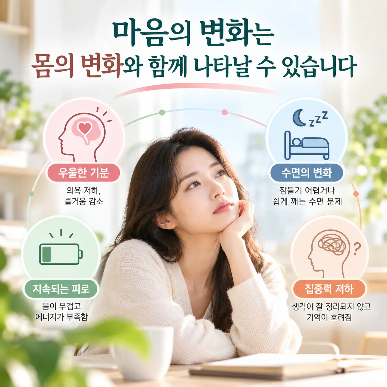
    우울증에서는 지속적인 우울감, 흥미 감소, 무기력감, 불안감이 나타날 수 있다. 또한 집중력 저하, 기억력 저하, 결정 능력 감소, 부정적 사고, 죄책감 등이 동반되기도 한다.

    신체적으로는 피로감, 수면장애, 식욕 변화, 두통, 소화기 증상 등이 나타날 수 있으며, 이러한 변화는 일상생활과 사회적 기능에 영향을 미칠 수 있다.

    우울증 신경기전의 발생 원인

    우울증은 단일 원인으로 설명하기 어려운 복합 질환이다. 만성 스트레스, 대인관계 갈등, 상실 경험, 직장 및 학업 부담 등은 우울증 발생 위험을 높이는 요인으로 알려져 있다.

    또한 유전적 소인, 신경생물학적 변화, 수면 부족, 만성 피로, 신체 질환, 사회적 고립 등도 우울증과 관련될 수 있다.

    우울증 신경기전 연구에서는 세로토닌, 도파민, 노르에피네프린과 같은 신경전달물질뿐 아니라 스트레스 반응 체계와 뇌 회로의 기능 변화도 중요하게 다뤄진다. 최근에는 우울증 신경기전을 통해 감정 조절과 인지 기능, 동기 부여 시스템의 변화를 함께 이해하려는 연구가 이루어지고 있다.

    우울증 신경기전과 현대 의학적 이해

    우울증의 세로토닌(serotonin) 신경전달 시스템

    우울증 신경기전 중 세로토닌 신경전달 시스템을 설명하는 이미지

    세로토닌은 기분 조절, 수면, 식욕, 충동 조절에 관여하는 대표적인 신경전달물질이다. 우울증에서는 세로토닌 신호 전달 기능의 변화가 우울감과 불안감 증가에 영향을 미치는 것으로 설명된다.

    우울증의 도파민(dopamine) 보상 회로 변화

    우울증 신경기전 중 도파민 보상 회로 변화를 설명하는 이미지

    도파민은 동기와 보상 체계를 조절하는 신경전달물질이다. 우울증에서는 도파민 관련 보상 회로의 활성 저하가 나타날 수 있으며, 이로 인해 흥미 감소와 의욕 저하가 발생하는 것으로 알려져 있다.

    노르에피네프린(norepinephrine)과 각성 조절

    노르에피네프린은 집중력과 각성 상태를 조절하는 역할을 한다. 기능 변화가 발생하면 무기력감, 피로감, 집중력 저하가 나타날 수 있다.

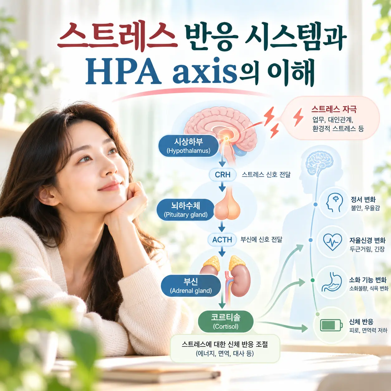
    우울증의 스트레스 반응 시스템과 HPA axis

    우울증 신경기전과 HPA axis 스트레스 반응 시스템을 설명하는 이미지

    만성 스트레스는 시상하부-뇌하수체-부신축인 HPA axis의 과도한 활성과 관련될 수 있다. 이러한 변화는 코르티솔 분비 이상을 유발하며 정서 조절 기능에 영향을 줄 수 있다.

    이러한 신경생물학적 변화는 한의학에서 설명하는 간기울결, 심비양허, 기혈양허와 기능적으로 연결되는 측면이 있다고 해석되기도 한다.

    우울증 신경기전의 한의학적 병리 이해

    우울증과 관련된 뇌 기능 변화를 시각화한 한국인 모델 이미지

    한의학에서는 이러한 상태를 정서 변화와 신체 기능 균형의 관점에서 설명한다. 특히 스트레스 반응, 신경계 긴장, 자율신경 균형 변화가 신체 기능과 정서 반응에 영향을 미치는 과정으로 해석한다.

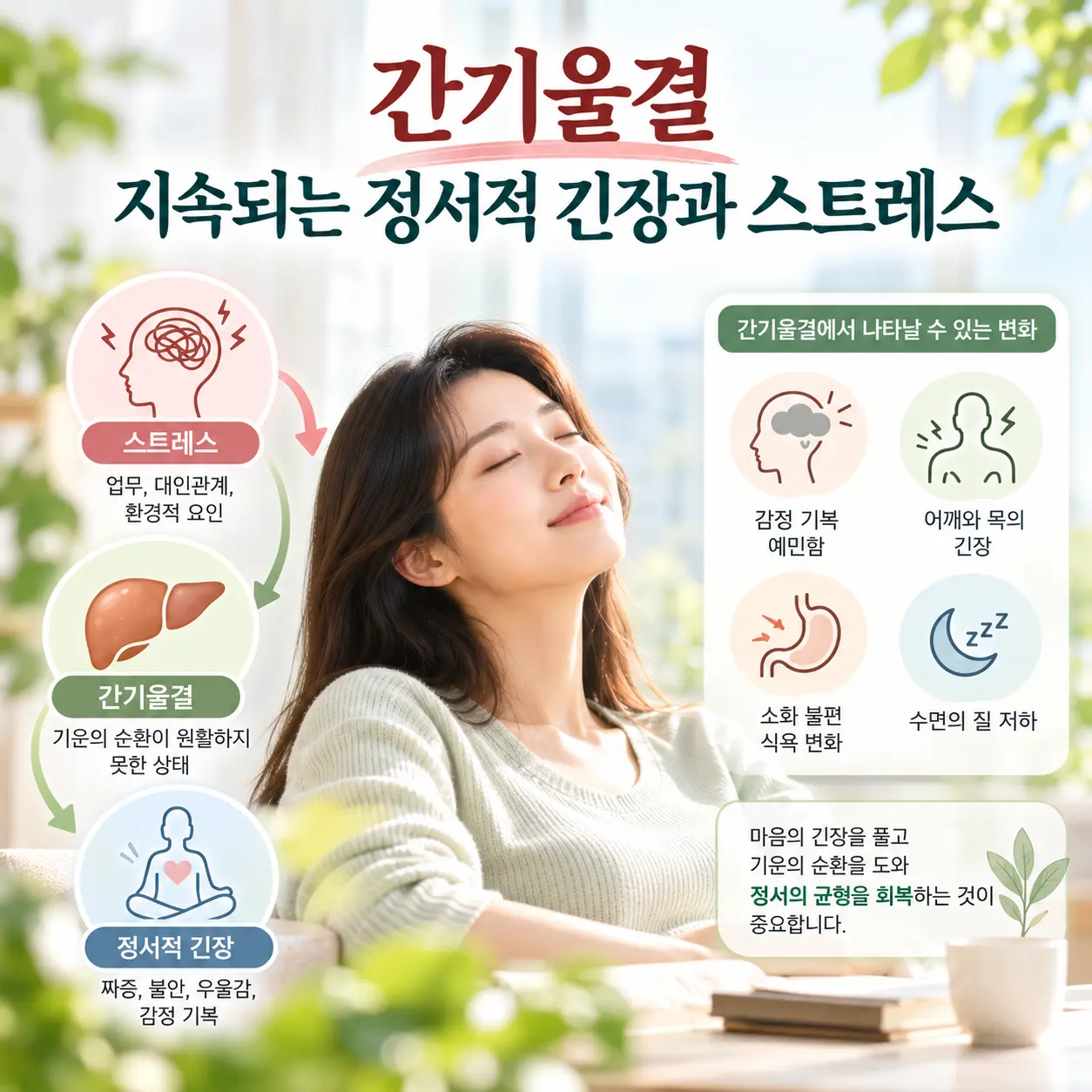
    간기울결(肝氣鬱結)과 정서 조절 변화

    간기울결은 스트레스나 감정 억압으로 인해 기의 순환이 원활하지 못한 상태를 의미한다. 정서적 긴장이 지속되면 신경계의 스트레스 반응이 증가하고 가슴 답답함, 우울감, 불안감 등이 나타날 수 있다고 설명한다.

    우울증에서 흔히 관찰되는 정서 위축과 의욕 저하는 간기울결과 관련하여 해석되기도 한다.

    심비양허(心脾兩虛)와 정신적 피로

    심비양허는 과도한 스트레스와 정신적 소모가 지속되면서 정신 활동과 신체 에너지 유지 기능이 저하된 상태를 의미한다. 심비양허가 나타나면 집중력 저하, 기억력 저하, 피로감, 수면장애 등이 동반될 수 있다.

    기혈양허(氣血兩虛)와 에너지 저하

    기혈양허는 신체를 유지하는 에너지와 영양 공급 기능이 부족한 상태를 의미한다. 기혈양허가 지속되면 무기력감, 쉽게 피로해지는 증상, 의욕 저하 등이 나타날 수 있으며 우울증의 신체적 쇠약감과 기능적으로 연결하여 이해하기도 한다.

    이처럼 간기울결, 심비양허, 기혈양허는 각각 독립적인 개념이라기보다 스트레스 반응, 신경계 조절 변화, 정서 기능 저하와 연관된 병리 과정으로 해석된다.

    우울증 신경기전의 한의학적 치료 접근

    우울증과 연관된 수면 문제 및 피로감을 표현한 한국인 성인 모델

    한의학 치료에서는 신경계 안정과 자율신경 균형 회복을 중요한 치료 목표로 설명한다.

    우울증 관리에서는 과도한 스트레스 반응을 완화하고 신체 회복력을 높이는 방향의 접근이 활용될 수 있다. 침 치료와 한약 치료 등 한의학적 치료 방법이 활용될 수 있으며, 정서 조절 기능과 수면 상태, 신체 컨디션 회복을 함께 고려한다.

    신경계 균형 회복과 정서 안정을 상징하는 한국인 성인 모델

    또한 생활 습관 관리와 상담적 접근을 병행하여 신경계 안정, 자율신경 조절, 기혈 순환 개선 및 신체 회복 과정을 돕는 방향으로 설명된다.

    정리

    우울증 신경기전은 세로토닌, 도파민, 노르에피네프린 등의 신경전달물질 변화와 뇌 기능 변화, 스트레스 반응 체계 이상을 포함하는 복합적인 과정으로 설명된다.

    한방신경정신의학에서는 이러한 변화를 정서와 신체 기능의 균형이라는 관점에서 이해하며, 간기울결, 심비양허, 기혈양허 등의 병리 개념을 통해 신경계 변화와 정서 반응을 함께 해석한다.

    함께 읽어보면 좋은 글

    우울증 증상과 진단 기준
    우울증 원인과 스트레스 반응
    우울증 치료와 관리 방법

    최근 우울증 신경기전에 대한 연구는 뇌 기능 네트워크와 신경전달물질의 상호작용을 중심으로 확대되고 있다. 특히 우울증 신경기전을 이해하는 것은 증상 발생 과정을 파악하고 다양한 치료 접근을 이해하는 데 도움이 될 수 있다.

    우울증에 대한 추가적인 정신건강 정보는 국립정신건강센터에서 확인할 수 있다.